
The human urinary system is a complex biological renal system that filters blood and excretes the unnecessary toxic waste components in the form of urine.
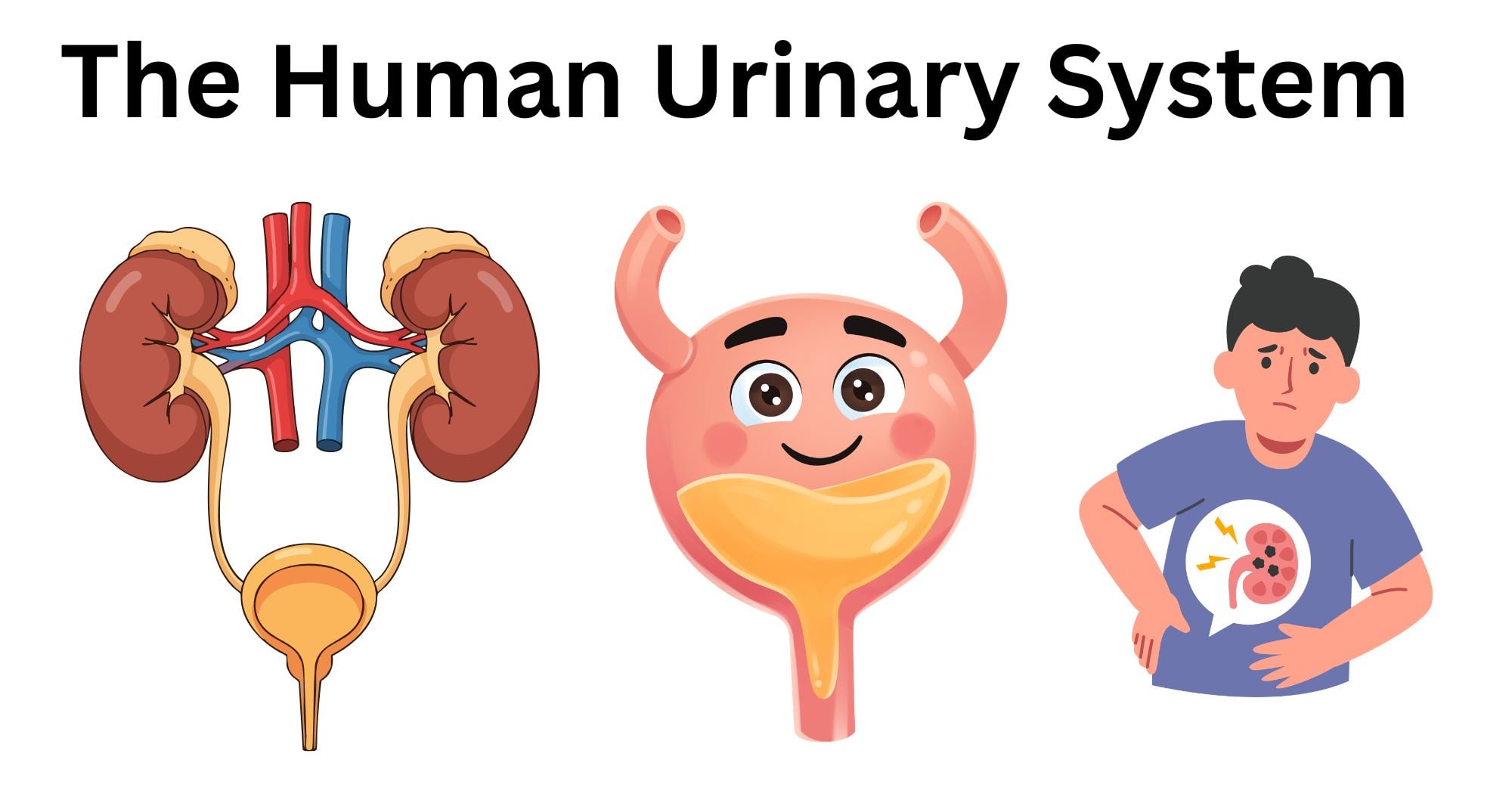
It also plays a significant role in maintaining the pH of blood, regulating its composition, volume, and pressure, and monitoring the body’s chemical-water balance.
Structure and Organs of the Human Urinary System
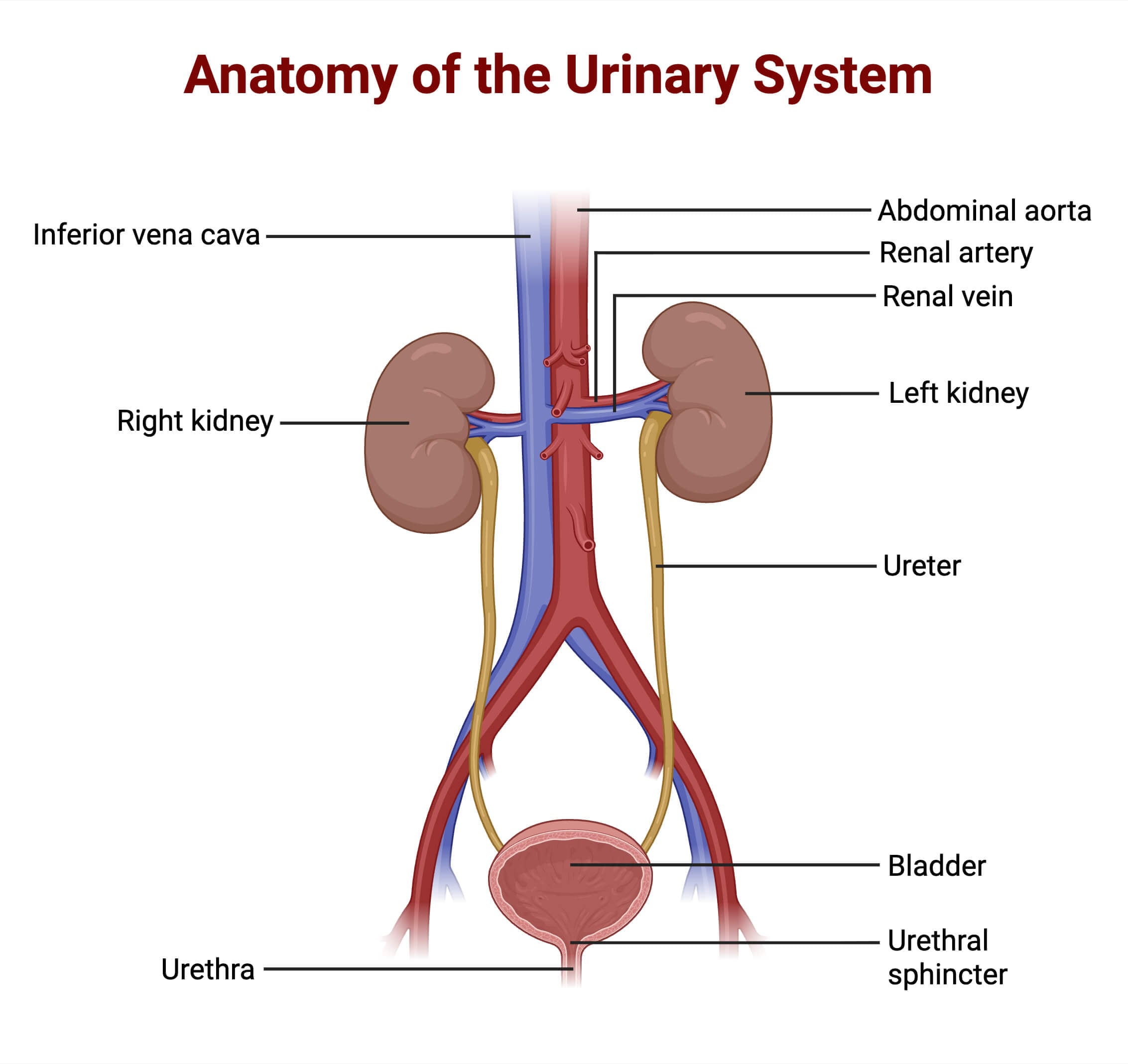
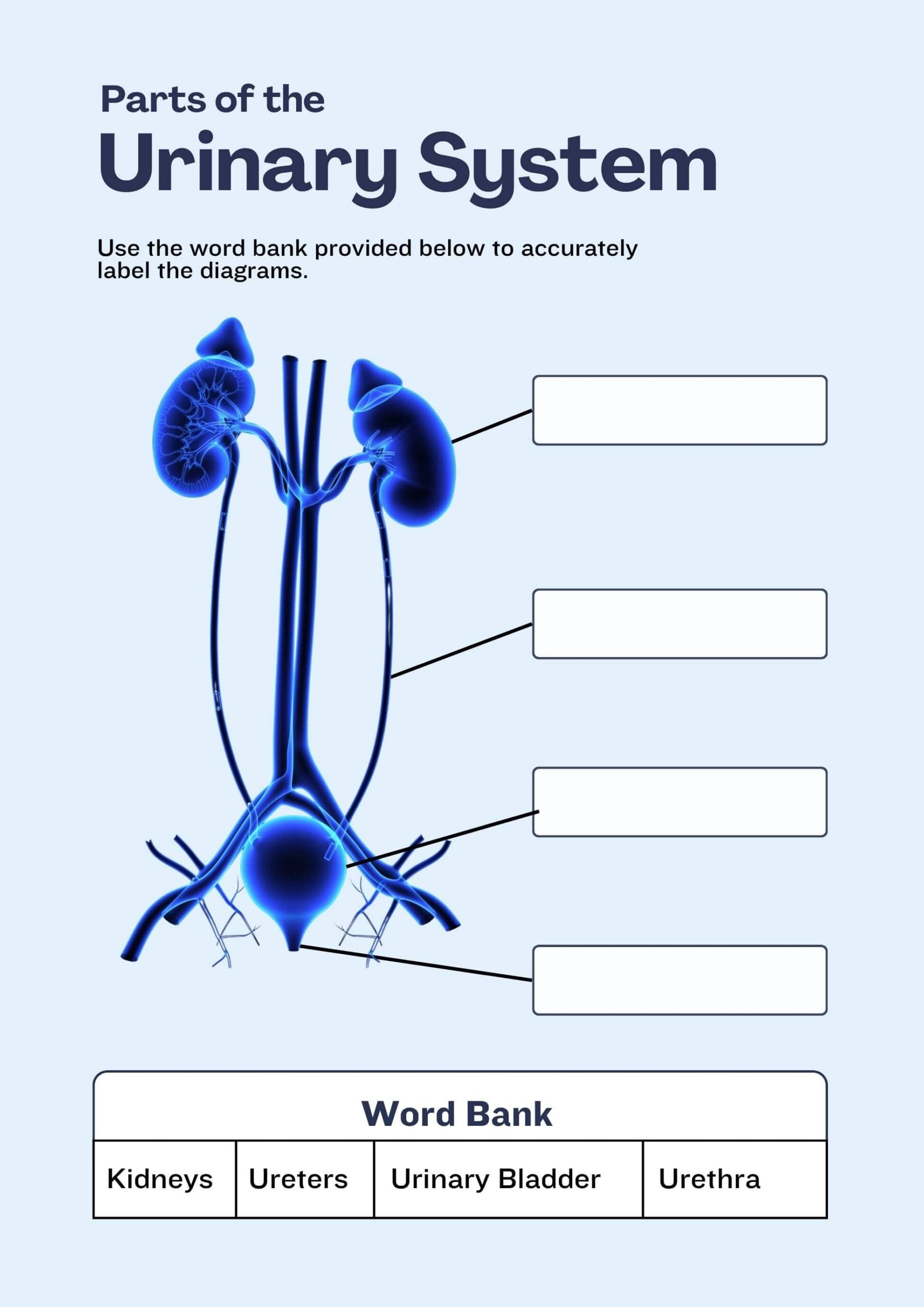
The human urinary system comprises the kidneys, Ureters, Urinary bladder, Urethra, and urinary sphincter muscles.
Kidneys
- Kidneys are bean-shaped organs present in a pair, inside the retroperitoneal space in the human body.
- They are the primary organs of the human excretory system along with the human urinary system.
- The right kidney is located slightly lower than the left kidney.
- They produce hormones, filter the blood in a continuous pattern, and separate out toxic components from the circulatory system.
Ureters
- They are the tubes extended from the kidneys to the urinary bladder.
- They are made up of smooth muscles that function to transport the urine to the bladder.
Urinary Bladder
- It is the hollow dome-shaped organ that is located in the lower abdomen, inside the pelvic region.
- It stores the urine temporarily.
- When the internal muscles of the bladder contract, the urine is eliminated via the urethra.
- Normally, an adult body can hold 300-600ml of urine in the bladder.
Urethra
- It is the muscular tube that is connected to the urinary bladder.
- It eliminates the urine from the body.
- The length of the urethra in a human male is nearly 18-20 cm, as the urine passes through the penis, while that in a human female is 4 cm.
- In females, the urine passes through the external opening between the clitoris and the vaginal opening.
Urinary sphincter muscles
- They are also called the urethral muscles.
- There are two types of sphincter muscles: internal and external.
- Internal sphincter muscles are made up of smooth muscles. Thus, they are involuntary, while external urethral muscles are made up of voluntary skeletal muscles.
- These muscles contract and relax during micturation (urination).
Functions of the Human Urinary System
- The urinary system helps to remove toxic waste components from the body.
- It helps to balance the fluids (water and chemicals) in the body.
- It also regulates the body’s pressure by maintaining the level of sodium in the blood.
- A proper balance of the ions and minerals, like Na+, K+, Ca++, Mg++, etc., is essential for the regulation of the body. The urinary system plays a major role in balancing these ions in the body, which is also crucial for a healthy life.
Anatomy of the Kidney
- Kidneys are a pair of bean-shaped internal organs that play a significant role in blood filtration.
- They are present in the abdominal cavity, located one on either side of the spine.
- Each kidney is about 10-12 cm long, 5-7cm wide, 2-3cm thick, and weighs about 120-170 gm.
- The right kidney is comparatively smaller and is also located at a lower level than the left kidney.
- They are covered and protected by the outermost capsule of fat and muscles, which is called the perirenal fat.
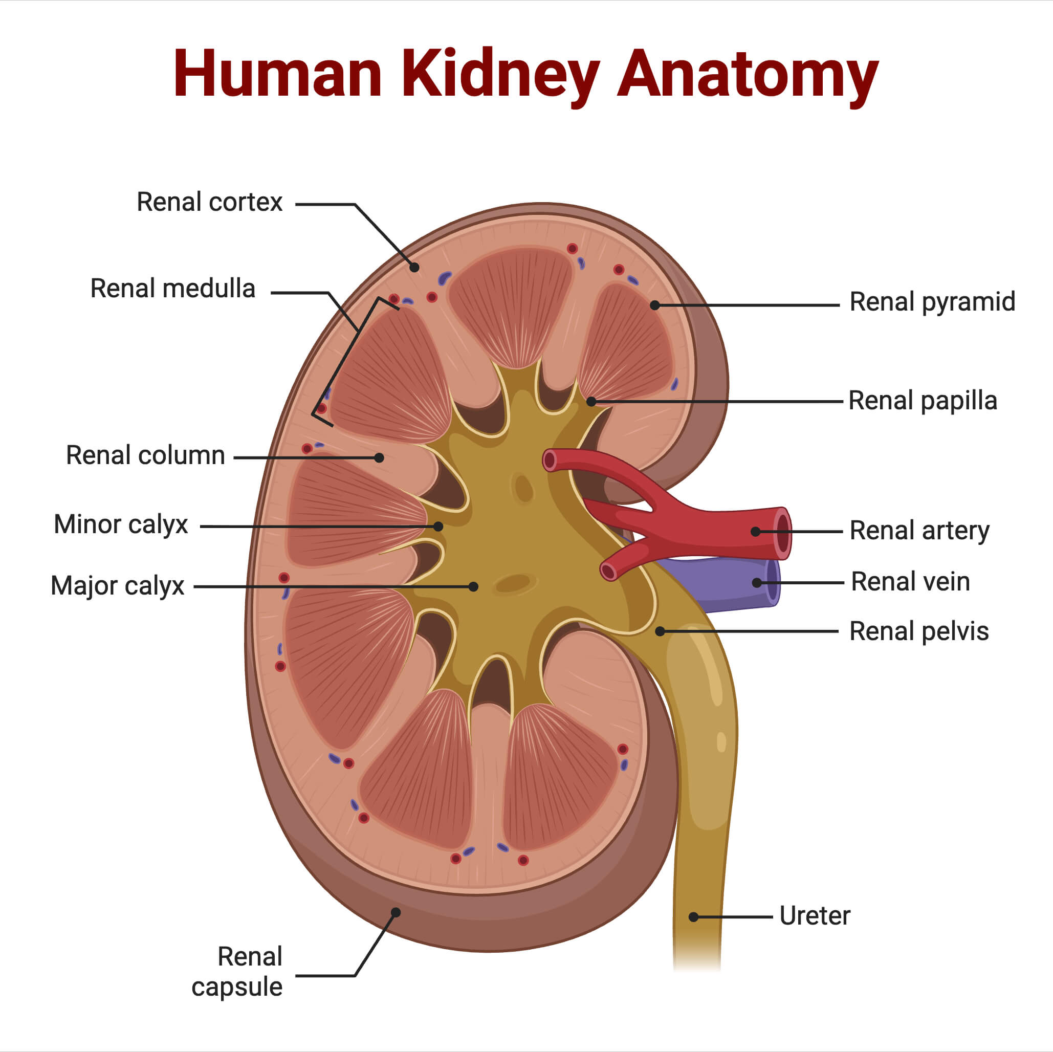
- Each human kidney is primarily divided into two structures: The renal cortex and the renal medulla
- Renal veins, arteries, and nerves, along with the ureter, enter and exit through the kidney via the renal hilum. The renal hilum is the fissure-like structure that is present in the renal pelvis. The renal pelvis is the funnel-shaped proximal part of the ureter in the kidney.
- The renal cortex is the outermost part of the kidney that covers the inner medulla. They control and regulate the balance of fluid and electrolytes.
- They are the vascular part of the kidney, which contains the renal corpuscles and renal tubules, apart from the region of Henle’s loop in the nephron.
- The functional unit of the kidney, nephrons, also begin in the renal cortex and extend up to the medulla.
- The renal cortex helps in the formation of red blood cells in the body by producing the erythropoietin hormone.
- The renal medulla is the inner part of the kidney that is divided into specific cone-shaped tissues, called the renal pyramids, projecting out as major and minor calyces. The human body contains approximately 10 -18 renal pyramids.
- The renal medulla also contains collecting ducts and the loop of Henle, the parts of nephrons that are responsible for maintaining the ionic balance in the blood.
Anatomy of Nephrons
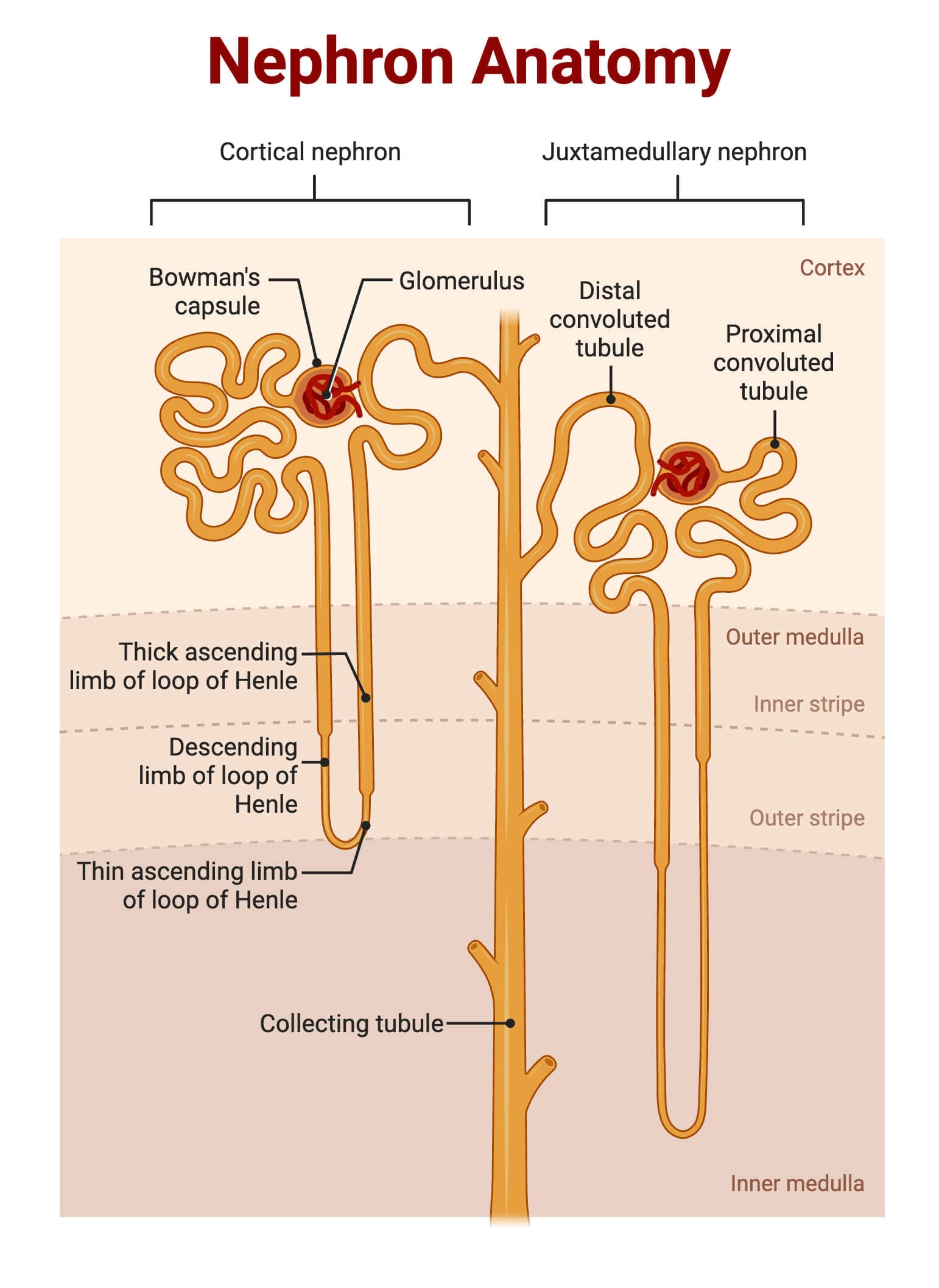
- Nephrons are the functional unit of the kidney. There are approximately over 1 million nephrons in the kidney.
- They consist of the following structures:
Renal corpuscles
They are the filtering units of the kidney. They consist of :
- Glomerulus: It is the network of capillaries present inside the Bowman’s capsule. As the blood enters the kidney via the afferent arterioles, the glomerulus filters the water and solutes from nearly one-fifth of the blood plasma and passes the remaining blood through the efferent arteriole.
- Bowman’s capsule: It is the double-layered cup-shaped structure surrounding the glomerulus. It is also called the glomerular capsule, which passes down the filtrate to the renal tubules.
Renal tubules
They are the tubes that carry the filtrate formed after blood filtration.
- Proximal convoluted tubules (PCT): They increase the surface area for absorption and are present in the renal cortex.
- Loop of Henle: It is the U-shaped tube that creates the concentration gradient in the renal medulla and regulates the ion reabsorption from the urine. The descending limb of the loop is permeable to water and has comparatively lower permeability for ions and urea. The ascending limb functions antagonistically to the descending limb and is more permeable to ions like Na+, K+, and Ca++, while being impermeable to water.
- Distal convoluted tubule (DCT): It is present between Henle’s loop and the collecting duct. It controls Na+ and K+ levels in the blood and also regulates the pH of the blood.
- Collecting duct: It is the collection of tubules that connect nephrons and the renal pelvis.
Formation of Urine and Process of Excretion
- When the blood flows through the kidney, the unnecessary ions and excess metabolic wastes are filtered in the glomerulus regions of the nephron.
- The filtrate is passed down the glomerulus and via the renal tubules. Different segments of the tubules are responsible for either absorption or secretion of water or electrolytes, which consequently determines the concentration of the filtrate.
- The filtrate that passes down the collecting ducts of the nephron, containing the unnecessary toxic metabolites such as urea, creatinine, ammonia, etc., is called urine.
- The urine formed in the nephron flows through the ureter. It is stored in the urinary bladder temporarily, and once the bladder gets full, the urinary sphincter muscles get activated, and they eliminate the urine via the urethra.
Steps and Mechanism of Urine Formation
Urine is composed of 95% water and the remaining 5% metabolic wastes, such as urea, creatinine, ammonia, and salts. It is passed down to the urinary bladder through the ureters and is excreted out of the body via the urethra.
It is formed in the nephrons by 3 major processes: Filtration, Reabsorption, and Secretion.
Filtration
- It is the first step in urine formation performed by the glomerulus.
- Blood circulates throughout the body and carries essential nutrients, including harmful toxic wastes. The kidneys filter nearly 1.1L to 1.2L of blood per minute.
- The efferent arteriole of the glomerulus is comparatively narrower than the afferent arteriole. This creates a pressure gradient known as glomerulus hydrostatic pressure. It filters the blood through the epithelium layer of Bowman’s capsule, the endothelium layer of glomerular blood vessels, and the basement membrane between these two layers.
- When the blood in the afferent arteriole enters the glomerulus, the glomerular hydrostatic force and the slit pores present in the Bowman’s capsule filter all the components of blood plasma, except the proteins.
- The volume of blood filtered from the kidney per unit time is called the Glomerular Filtration Rate (GFR). It is approximately 180L per day.
Reabsorption
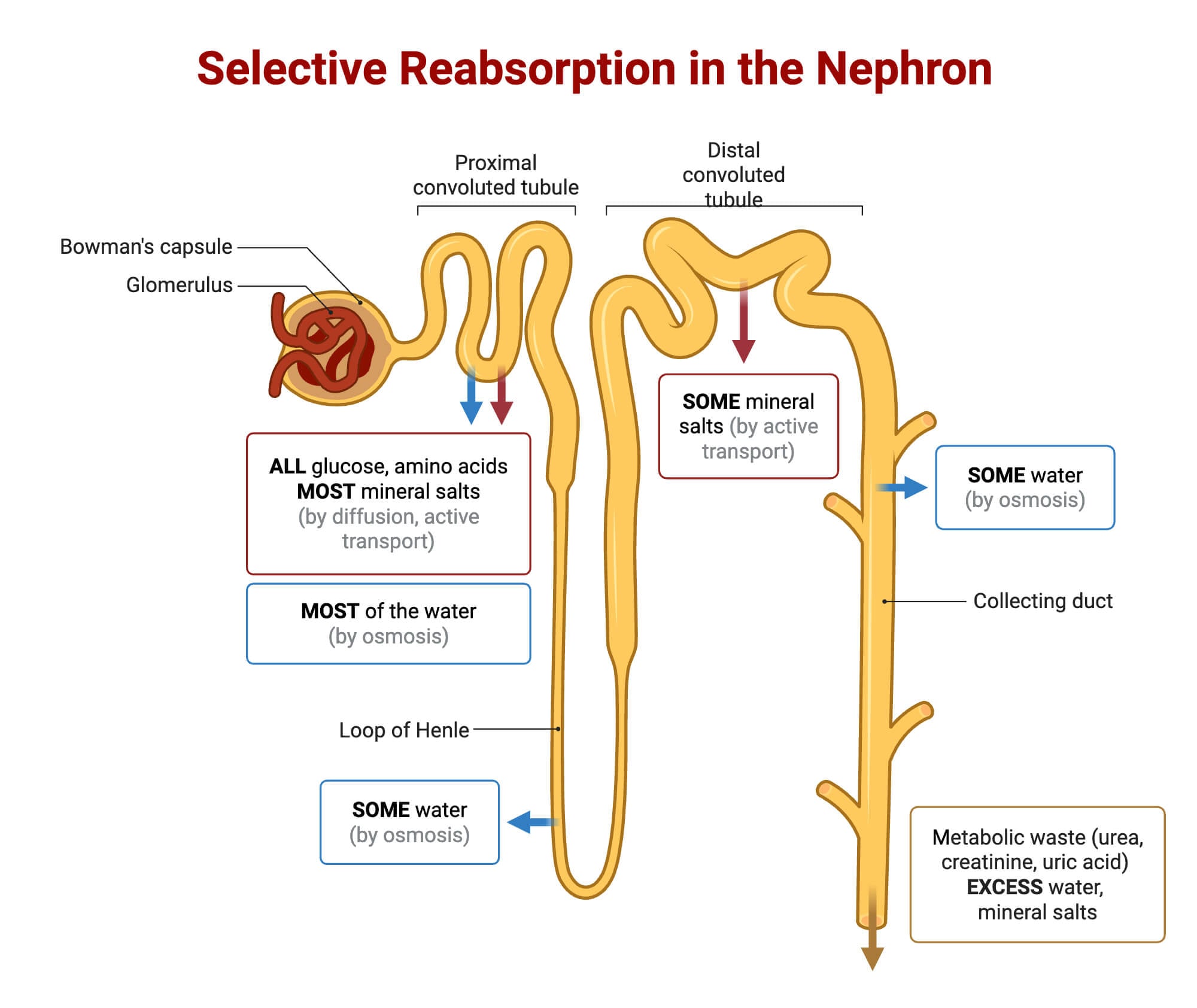
- In the renal tubules, the blood, filtered through the glomerulus, is selectively reabsorbed by either active or passive transport mechanisms.
- Different kinds of ions and valuable substances are absorbed by the blood capillaries present in the proximal convoluted tubule.
- PCT reabsorbs 55% of water. They reabsorb the essential nutrients and ions such as glucose, sodium, amino acids, lactate, etc from the filtrate by active transport, while nitrogenous wastes and water are reabsorbed by passive transport.
- Henle’s loop also plays a minor yet significant role in reabsorption. The descending limb of the loop is highly permeable to water, while the ascending limb is permeable to ions. Due to this, the body fluid is highly concentrated with ions and electrolytes in the descending limb. As the fluid passes to the ascending limb, the concentration of the fluid decreases, and the fluid gets diluted.
- Distal convoluted tubules also reabsorb the bicarbonate ions.
- The collecting duct reabsorbs a large volume of water from the filtrate and produces concentrated urine.
Secretion
Another important step of urine formation is secretion. Renal tubules play a major role in maintaining ionic balance. They secrete ions like H+, K+, and NH3+ ions and release a large amount of water.
Regulation of fluid, electrolyte, and pH balance
- The kidneys play a major role in maintaining homeostasis in the body. It regulates the body fluids, electrolytes, and also controls the acid-base balance(pH) in the blood.
- It controls the volume of water and concentration of ions in the body.
- Regulation of body fluids and electrolytes: Anti-diuretic hormone (ADH) stimulates the kidneys for water reabsorption in the collecting ducts. When the amount of water is less in the body, ADH hormone stimulates the kidneys to reabsorb water, and thus, after blood filtration, concentrated urine passes through the ureter.
- The kidneys are also responsible for regulating the amount of electrolytes. When the body gets dehydrated, the amount of electrolytes, such as sodium, also decreases in the body. It is because the volume of water regulates the level of osmolarity (concentration of solutes/volume) in blood.
- Aldosterone hormone (secreted by the adrenal cortex) promotes the reabsorption of sodium ions in the cells from the filtrate in the PCT and DCT. Consequently, the potassium ions are excreted from the tubular cells into the urine via the sodium-potassium pump.
- Regulation of pH: The renal tubules in the kidneys selectively remove the excess amount of H+ ions and selectively reabsorb the bicarbonate ions. This process helps to prevent blood alkalosis and maintain the blood pH.
Regulating blood pressure and fluid balance
- The human body regulates blood pressure by secreting specific enzymes and hormones in the blood.
- As the blood pressure decreases, kidney secretes renin enzyme and activates renin- angiotensin-aldosterone-system (RAAS) and activate the vasoconstrictor effect(angiotensin-II). The blood vessel then narrows down and elevates the pressure within the vessel walls.
- Moreover, angiotensin-II also stimulates the secretion of aldosterone and helps to regulate the sodium and water reabsorption system in the kidney.
Male vs. Female Urinary System
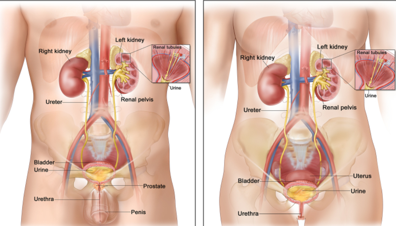
Source: NCBI Book.
| Features | Male Urinary System | Female Urinary System |
| Urethra | In males, the urethra is 18-20cm long and is essential for the ejaculation of semen along with urine excretion. It belongs to both the urinary and reproductive systems. | In females, the urethra is comparatively shorter than in males(4cm) and functions to eliminate urine. It only belongs to the urinary system. |
| Urinary Sphincter Muscles | They control the excretion of both semen and urine in males. | In females, they control the movement of urine only. |
| Urine path | curvy | straight |
| Catheterizations | difficult | not so difficult |
| Bacterial infection | Bacterial infections are rare. | Bacterial infection and UTIs are most common in females. |
| Capacity of the Urinary Bladder | A male bladder can hold up to 700ml of urine. | A female bladder can hold up to 500 ml of urine. |
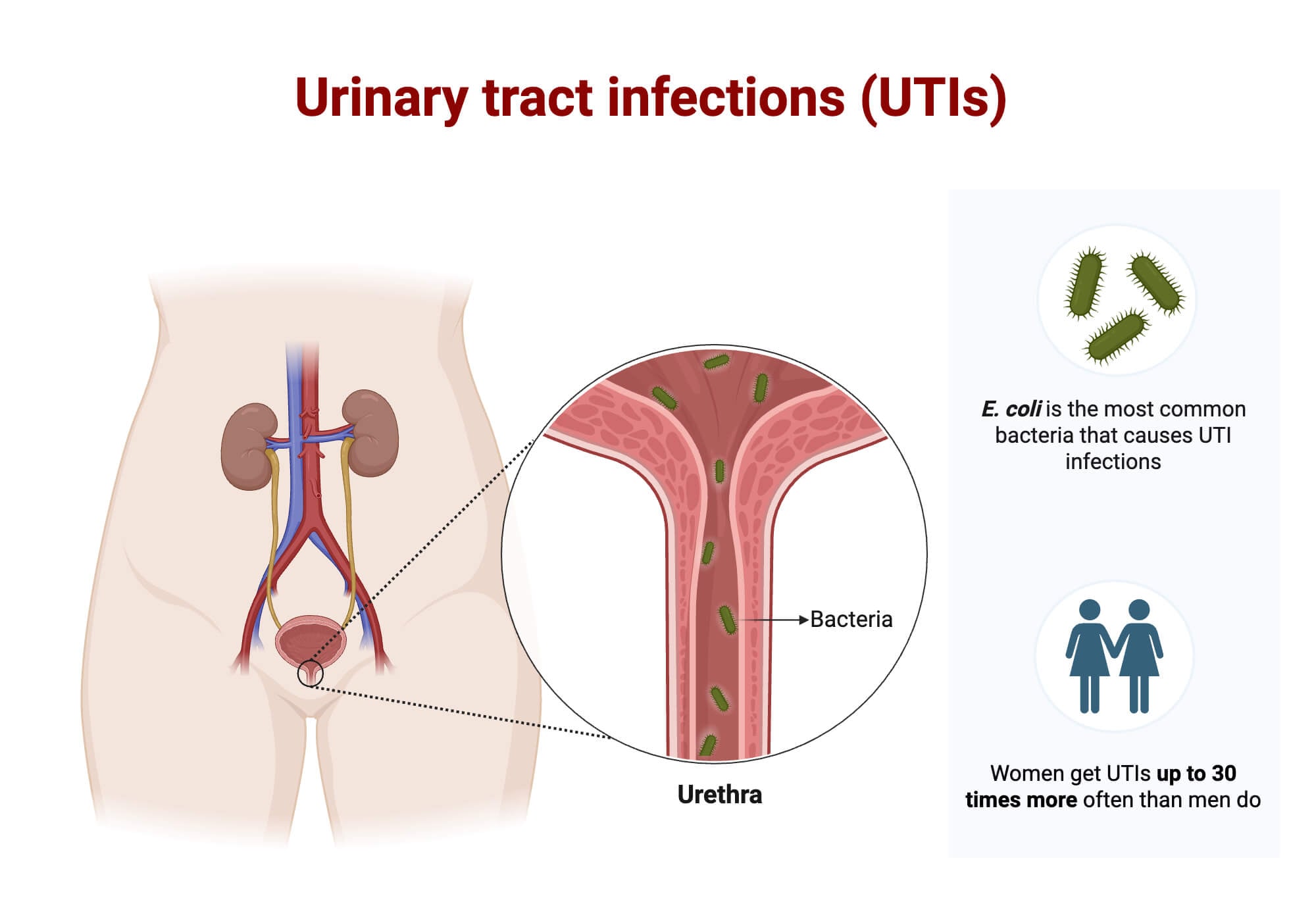
| UTIs | The risk of UTIs is comparatively lower in males due to their long urethra. | The risk of UTIs is higher in females because they have a shorter urethra |
Common Diseases and Disorders of the Human Urinary System
Urinary tract infections (UTIs) are the most common type of disease in humans. When the organs involved in urine formation and excretion become infected with bacteria, this causes UTIs. The most common types of UTIs are cystitis, urethritis, and Pyelonephritis.
| S.N | Diseases/ Disorders | Causes | Symptoms |
| 1. | Kidney Stones | It is caused by the accumulation of crystals formed by the waste materials present in the urine. | Severe pains, lower abdominal cramps, nausea, vomiting |
| 2. | Urethritis | It is caused by the viral or bacterial inflammations or urethra | In males: Pain or swelling of the penis, blood in urine or semen, frequent urination. In females: frequent urination, fever, chills, vaginal discharge |
| 3. | Cystitis (Bladder Inflammation) | It is used for the bacterial infection of the urinary bladder | Frequent urination, dysuria, and abdominal pain |
| 4. | Pyelonephritis | It is the inflammation of the kidney caused by the spread of bacteria to the urinary tract | Fever, chills, back pain, blood in urine, frequent urination |
| 5. | Chronic Kidney Disease (Kidney failure) | It is caused when the kidney fails to filter the blood. | Fatigue, decreased urination, nausea, vomiting |
How to Keep the Urinary System Healthy?
We can keep our urinary system healthy by following the given measures:
- Drinking plenty of water, fruit juices, and other fluids to prevent dehydration
- Discharge the urine once you feel like the bladder is full. Holding the urine for too long increases the risk of bladder infections.
- Regular physical exercise helps to prevent constipation
- Avoid caffeinated drinks, as it increases the urge for frequent urination
Conclusion
The urinary system in the human body is the biological system primarily responsible for removing harmful substances as urine. It also maintains homeostasis and regulates the concentration of water and electrolytes in the bodily fluids.
References
- 15 tips to keep your bladder healthy. (2022, January 24). National Institute on Aging. https://www.nia.nih.gov/health/bladder-health-and-incontinence/15-tips-keep-your-bladder-healthy
- Biga, L. M., Bronson, S., Dawson, S., Harwell, A., Hopkins, R., Kaufmann, J., LeMaster, M., Matern, P., Morrison-Graham, K., Oja, K., Quick, D., Runyeon, J., & OpenStax. (2025, September 1). 25.1 Internal and External Anatomy of the Kidney. Anatomy & Physiology 2e. https://open.oregonstate.education/anatomy2e/chapter/internal-external-anatomy-kidney/
- Breshears, M. A., & Confer, A. W. (2017). The Urinary System. Pathologic Basis of Veterinary Disease, 617–681.e1. https://doi.org/10.1016/B978-0-323-35775-3.00011- 4
- Contributors to Wikimedia projects. (2004, June 9). Human Physiology/The Urinary System. Wikibooks, Open Books for an Open World. https://en.wikibooks.org/wiki/Human_Physiology/The_Urinary_System#References
- Coronal section of the kidney. (2023, October 30). Kenhub. https://www.kenhub.com/en/library/anatomy/coronal-section-of-the-kidney
- ERA. (2023, October 10). Understanding kidneys, Facts and Functions | ERA. https://www.era-online.org/publications/understanding-kidneys/#:~:text=The%20glomerular%20filtration%20rate%20(GFR,important%20for%20examining%20kidney%20function.
- Fluid and electrolyte balance. (n.d.). https://mcb.berkeley.edu/courses/mcb135e/kidneyfluid.html
- Gantsova, E., Serova, O., Vishnyakova, P., Deyev, I., Elchaninov, A., & Fatkhudinov, T. (2024). Mechanisms and physiological relevance of acid-base exchange in functional units of the kidney. PeerJ, 12, e17316. https://doi.org/10.7717/peerj.17316
- Hague, Andrew. (2018). What goes in, must come out. General Medicine Open. 2. 10.15761/GMC.1000140.
- “Kidneys: Anatomy, Function, Health & Conditions”. Cleveland Clinic. Archived from the original on 2023-06-29. Retrieved 2023-07-13.
- Lepowsky, E., Ghaderinezhad, F., Knowlton, S., & Tasoglu, S. (2017). Paper-based assays for urine analysis. Biomicrofluidics, 11(5), 051501. https://doi.org/10.1063/1.4996768
- Libretexts. (2024, December 24). 26.2: Functions of the Urinary System. Medicine LibreTexts. https://med.libretexts.org/Bookshelves/Nursing/Fundamentals_of_Nursing_(OpenStax)/26:_Urinary_Elimination/26.02:_Functions_of_the_Urinary_System
- News-Medical. (2018, October 24). Anatomy of the kidney. https://www.news-medical.net/health/Anatomy-of-the-Kidney.aspx
- Ogobuiro I, Tuma F. Physiology, Renal. [Updated 2023 Jul 24]. In: StatPearls [Internet]. Treasure Island (FL): StatPearls Publishing; 2026 Jan-. Available from: https://www.ncbi.nlm.nih.gov/books/NBK538339/
- Professional, C. C. M. (2026, January 8). Urinary system. Cleveland Clinic. https://my.clevelandclinic.org/health/body/21197-urinary-system#function
- Standring, Susan, ed. (2016). “Bladder, prostate and urethra”. Gray’s Anatomy: The Anatomical Basis of Clinical Practice (41st ed.). Philadelphia: Elsevier Limited. pp. 1261–1266. ISBN 978-0-7020-5230-9. OCLC 920806541.
- Unknown. (n.d.-a). Physiology of urine formation. https://www.miperknlapindia.ac.in/urine-formation.pdf
- Unknown, U. (n.d.-b). Anatomy Lecture Notes Section 6: The Renal (Urinary) System. https://sdmiramar.edu/sites/default/files/2024-05/%2313%20Renal%20System%20Lect%20Notes.pdf
- Vaidya SR, Aeddula NR. Chronic Kidney Disease. [Updated 2024 Jul 31]. In: StatPearls [Internet]. Treasure Island (FL): StatPearls Publishing; 2026 Jan-. Available from: https://www.ncbi.nlm.nih.gov/books/NBK535404/
- Visible Body, part of Cengage Learning. (n.d.). Urinary system structures. https://www.visiblebody.com/learn/urinary/urinary-system-structures#:~:text=In%20males%2C%20the%20urethra%20is,the%20body%20through%20the%20urethra.
- Welcome to ToxTutor. (n.d.). Toxicology MSDT. https://www.toxmsdt.com/132-urinary-excretion.html
