
An ultrasound machine is a diagnostic imaging device that generates images of the internal body using high-frequency sound waves, called ultrasound.

What is an Ultrasound Machine?
An ultrasound machine, also called a sonography unit, is a medical device that uses high-frequency sound waves in the range of 2 – 18 megahertz (MHz) to produce an image of the internal body structures. It does not use ionizing radiation as done by X-ray or CT scanning, making it a safe imaging technique. The ultrasound machine generates images by emitting sound waves, which are bounced back from internal structures of the body, and a computer captures these reflections/ echoes and constructs an image. It is widely used in cardiology, abdominal imaging, musculoskeletal assessment, obstetrics, and so on for its portability, safety, and real-time imaging capability.
Principle of Ultrasound Imaging
Ultrasound imaging operates on the principle of echo reflection. It is analogous to sonar navigation. When an electrical voltage is applied to a piezoelectric crystal inside the transducer, the crystal vibrates and produces high-frequency sound waves. These waves travel through the body’s tissues and encounter different structures with different densities, such as the boundary between muscle and bone, or fluid and soft tissue. At each of these interfaces, a sound wave is reflected or bounced back, which is received by the transducer as echoes. The machine calculates the depth of each reflecting structure based on the time delay between the transmitted pulse and the received echo, using the known average speed of sound in soft tissue i.e., approx. 1,540 m/s. The machine then uses this to create a real-time image where the intensity of the reflected echo determines the brightness of the pixel in the image.
Simply, ultrasound imaging is based on the reflection of high-frequency sound waves from body tissues. A transducer transmits sound waves into the body, and the sound waves are bounced back when they encounter tissues and organs, which are received by the transducer. The ultrasound machine uses the strength of the echoes and the time taken for them to return to create images of internal body structures.
Components and Parts of an Ultrasound Machine
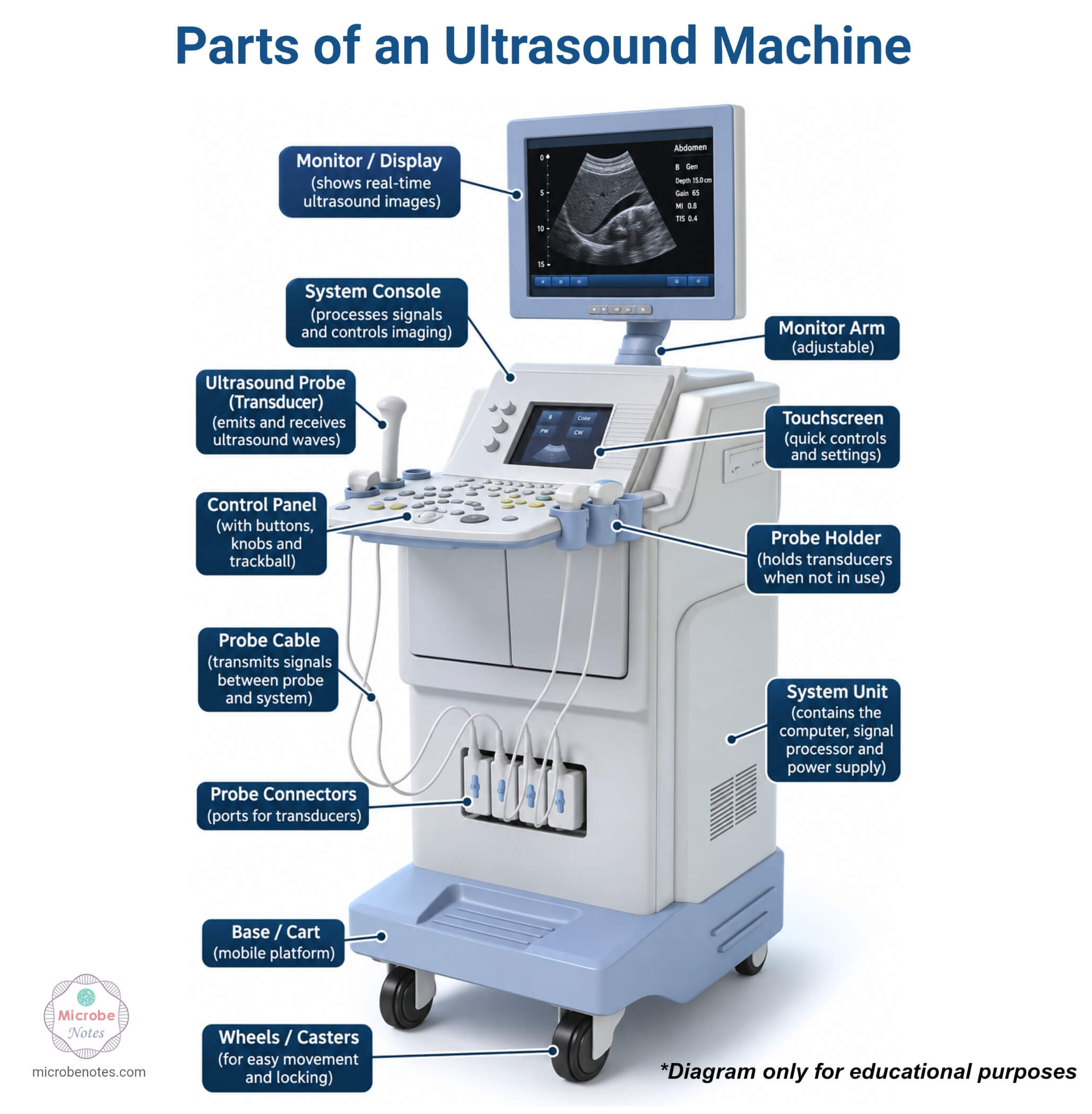
An ultrasound machine consists of core components, including a transducer, transmitter pulse generator, compensating amplifiers, control unit, digital processors, and display system. It combines two fundamental technologies, i.e., image production and Doppler assessment (Continuous wave, pulse wave, and color-flow mapping). The main components with functions are:
- Transducer or probe: generates and receives sound waves via piezoelectric crystals
- Pulse generator: produces strong, short electrical pulses to excite the transducer
- Compensating amplifiers: amplify returning echo signals for processing
- Digital processors: convert electrical signals into image data
- Display system: presents the constructed image to the operator
- Control unit: allows the operator to adjust settings such as depth, gain, frequency, etc.
Types of Ultrasound Machines
Ultrasound machines are broadly classified into the following types based on their design, portability, and application:
Cart-based/ Trolley ultrasound system: They are large ultrasound machines found in hospitals and radiology departments. They have advanced imaging capabilities.
Portable/ compact ultrasound machines: They are smaller, laptop-style systems used for bedside or field examination. They offer portability with slightly reduced image resolution as compared to a cart-based system.
Handheld ultrasound devices: They are pocket-sized devices that are connected to phones or computers. They are mostly used in point-of-care ultrasound (POCUS) settings, emergencies, and rural healthcare.
Specialized systems: They are machines designed for specific applications such as echocardiography, obstetric ultrasound, or musculoskeletal ultrasound. They often have dedicated software and transducers for the target organ system.
Types of Ultrasound Transducers (Probes)
There are a variety of probes available for scanning different parts of the body that are selected based on the size and shape of the beam. The choice of transducer affects the image quality and clinical application significantly.
Linear array transducer: produces high-frequency, high-resolution images and is commonly used for superficial structures like thyroid, vascular, and musculoskeletal imaging.
Curvilinear (convex) transducer: operates at lower frequencies with a wider field of view. It is ideal for abdominal and obstetric imaging.
Phased array transducer: It is commonly used in cardiac imaging to image through the intercostal spaces.
Endocavity transducer: It is designed for internal examinations such as transvaginal or transrectal ultrasound.
How Does an Ultrasound Machine Work?
The ultrasound machine operates by generating strong, short electrical pulses that excite the transducer to emit a focused sound wave into the body. The sound wave reflects off tissue boundaries with different acoustic resistance, and the returning echoes are captured by the same transducer and converted into electrical signals. These signals are amplified, processed by digital processors, and displayed as an image. Additional processing algorithms are often applied to enhance image clarity and reduce artifacts. The stability and repeatability of the ultrasound image can be improved by controlling the stability of the contact force.
Common Ultrasound Imaging Modes
Imaging can be performed in several modes, including A mode, B mode, M mode, and D mode. Different imaging modes serve different diagnostic purposes.
- A-mode (Amplitude mode): It displays echo amplitude as spikes along a single line, used in ophthalmology for axial length measurement.
- B-mode (Brightness mode): It produces a 2-D grayscale cross-sectional image based on echo intensity.
- M-mode (Motion mode): It displays the movement of the structure over time along a single scan line.
- D-mode (Doppler mode): It utilizes the Doppler effect to assess blood flow direction, velocity, and pattern.
- 3D/ 4D mode: It reconstructs volumetric data into 3D images and 4D mode.
- Elastography: It assesses tissue stiffness.
What are the applications of an Ultrasound Machine?
An ultrasound machine is used across many medical specialties. The applications are:
- In obstetrics and gynecology, they monitor fetal development, estimates gestational age, and detects congenital anomalies.
- In cardiology, echocardiography evaluates heart structure, valve function, and blood flow dynamics.
- Abdominal ultrasound assesses organs such as the liver, gallbladder, kidneys, and pancreas for abnormalities.
- Vascular ultrasound detects blood clots, arterial stenosis, and venous insufficiency.
- Musculoskeletal ultrasound evaluates tendons, ligaments, and joints.
- In the intensive care unit, it is a life-saving tool for critical care assessment.
Advantages of an Ultrasound Machine
- An ultrasound machine does not use ionizing radiation, making it safe for pregnant women and children.
- It offers real-time imaging.
- It is more cost-effective and portable than CT and MRI in many clinical situations.
- The procedure is non-invasive, painless, and does not require sedation.
- It enables repeated examinations without patient harm due to the absence of radiation.
Limitations of an Ultrasound Machine
- Ultrasound examinations are highly operator-dependent, creating inter- and intra-operator variability.
- Operators require significant training and experience to acquire and interpret images accurately.
- Conventional ultrasound equipment is difficult to operate due to its complex structure, large size, and space requirements.
- Ultrasound waves are transmitted poorly through air and bone.
Safety Considerations and Maintenance of an Ultrasound Machine
Even though ultrasound is considered safe, unnecessary exposure must be minimized because of the thermal and mechanical bioeffects of ultrasound. Regular maintenance is important to ensure accuracy and equipment durability. Maintenance includes routine cleaning and disinfection of transducers between patients to prevent cross-contamination. It also includes periodic calibration checks, inspection of cables and connectors for wear and tear, and necessary software updates. Transducers should be handled and stored properly as they contain piezoelectric elements that can be damaged by drops or improper storage. Proper documentation and quality control checks should be done to help ensure image quality and accurate diagnosis.
Conclusion
The ultrasound machine is one of the most important and advanced diagnostic medical devices. They have developed from large stationary systems to small, portable, and wearable systems over time and are essential tools in modern medicine. They are preferred because they do not use ionizing radiation, so they are safer for repeated exposure. It produces a real-time image and is portable, so it is a standard diagnostic test across nearly all organ systems.
It has diverse applications from obstetrics to cardiology and emergency medicine. So it is important to understand its components, working principle, and proper maintenance for professionals in medical sciences.
References
- Nayak, G., Bolla, V., Balivada, S. K., & P., P. (2022). Technological Evolution of Ultrasound Devices: A Review. International Journal of Health Technology and Innovation, 1(03), 24–32. https://doi.org/10.60142/ijhti.v1i03.55
- Carovac, A., Smajlovic, F., & Junuzovic, D. (2011). Application of Ultrasound in Medicine. Acta Informatica Medica, 19(3), 168. https://doi.org/10.5455/aim.2011.19.168-171
- Health, C. for D. and R. (2024). Ultrasound Imaging. FDA. https://www.fda.gov/radiation-emitting-products/medical-imaging/ultrasound-imaging
- Manual of diagnostic ultrasound. 2 (2. ed). (2013). World Health Organization.
- Vrdoljak, I., & Dravinac, L. (2024). The Role of Ultrasound in Medical Diagnostics and Treatment. ENTRENOVA – ENTerprise REsearch InNOVAtion, 10(1), 163–175. https://doi.org/10.54820/entrenova-2024-0015
- Lawrence, J. (2007). Physics and instrumentation of ultrasound. Critical Care Medicine, 35(8), S314-S322. https://doi.org/10.1097/01.CCM.0000270241.33075.60.
