
The magnetic bead-based DNA extraction is a modern and highly efficient technique widely used in molecular biology laboratories for isolating DNA from a variety of biological samples.
This method relies on the reversible binding of DNA to magnetic particles, typically coated with silica or other functional groups, under specific chemical conditions. Once bound, DNA can be easily separated from contaminants using an external magnetic field, eliminating the need for centrifugation steps.
The concept of using solid surfaces for nucleic acid purification has evolved significantly over time. Rohland and Hofreiter (2007) described early applications of silica-based extraction strategies, particularly for challenging samples such as ancient bones and teeth, where DNA is often highly fragmented and present in low quantities. The adaptation of this principle to magnetic beads has further improved efficiency, reproducibility, and scalability, making the method suitable for both research and clinical applications (Rohland & Hofreiter, 2007).
Magnetic bead-based DNA extraction is now commonly integrated into automated platforms, allowing high-throughput processing with minimal hands-on time. Due to its speed, reduced contamination risk, and compatibility with downstream applications such as PCR and sequencing, this method has become a preferred alternative to traditional phenol-chloroform and column-based extraction techniques (Brown, 2020).
Key Reagents of the Magnetic Bead-Based DNA Extraction
| Reagent | Typical Concentration / Amount | Purpose |
| Magnetic beads (silica-coated) | 10–50 µL per sample | Bind DNA selectively under chaotropic conditions |
| Lysis buffer | Variable (kit-specific) | Disrupt cell membranes and denature proteins |
| Proteinase K | ~20 mg/mL | Digest proteins and nucleases |
| Binding buffer (chaotropic salts) | High salt concentration | Promote DNA adsorption to beads |
| Wash buffer (ethanol-based) | 70–80% ethanol | Remove salts and contaminants |
| Elution buffer (Tris-HCl or water) | 50–100 µL | Release purified DNA from beads |
Principle of the Magnetic Bead-Based DNA Extraction
The fundamental principle of magnetic bead-based DNA extraction is derived from the silica-binding mechanism first described by Boom et al. (1990). Under high concentrations of chaotropic salts, such as guanidinium thiocyanate, hydrogen bonding between water molecules is disrupted. This promotes the adsorption of negatively charged DNA molecules onto silica surfaces.
In magnetic bead-based systems, silica is coated onto paramagnetic beads. When DNA binds to these beads, they can be rapidly separated from the solution using a magnetic field, while impurities remain in the supernatant. Subsequent washing steps remove residual contaminants, and DNA is finally eluted under low-salt conditions that destabilize the DNA-silica interaction (Boom et al., 1990).
This principle aligns with the broader silica-based purification strategies discussed in classical molecular biology texts, where solid-phase extraction is favored for its efficiency and reduced mechanical stress on DNA molecules (Brown, 2020).
Protocol of the Magnetic Bead-Based DNA Extraction
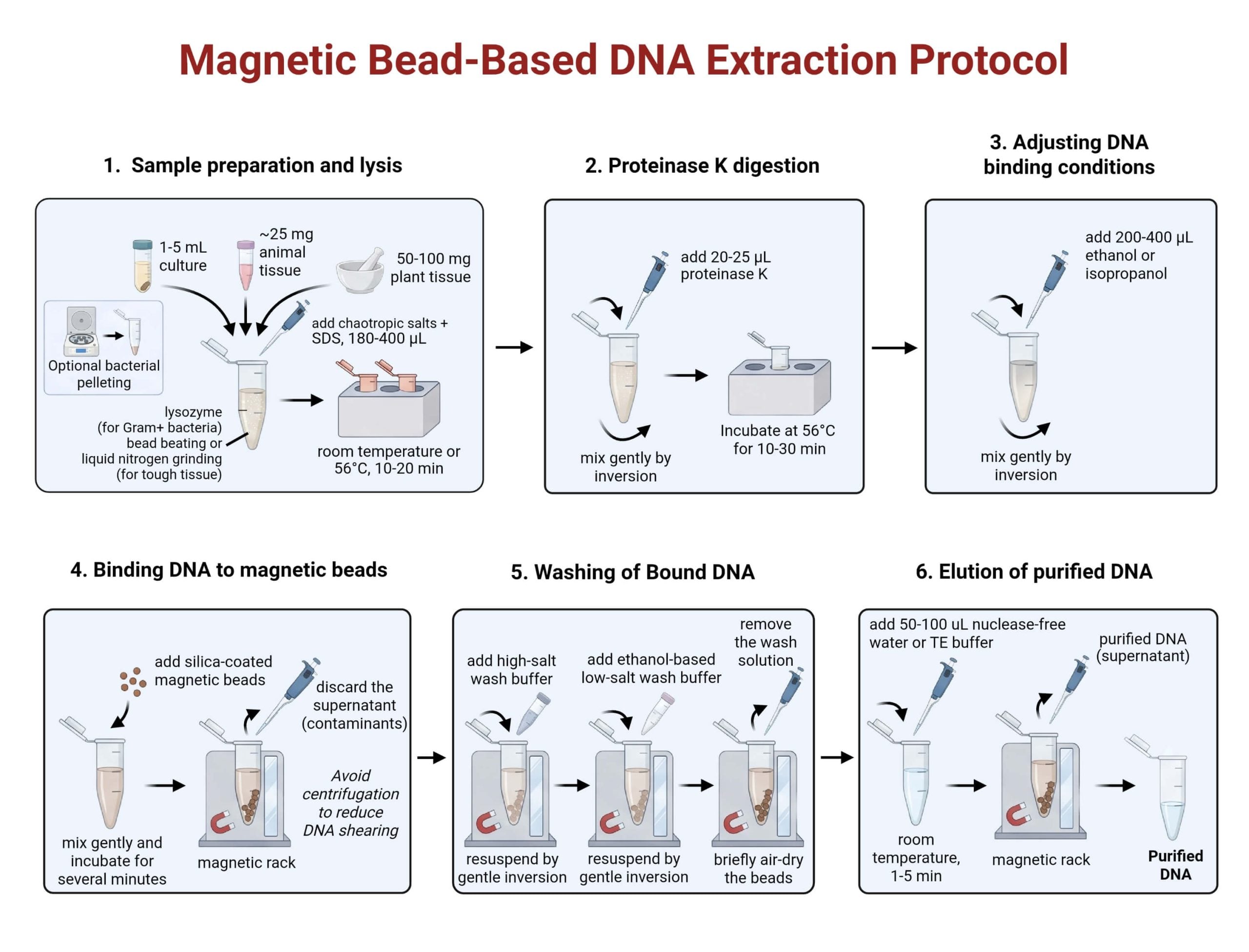
1. Sample Preparation and Lysis
- Collect the biological sample:
- Bacterial pellet: from 1–5 mL liquid culture
- Animal tissue: ~25 mg
- Plant tissue: 50–100 mg
- Transfer the sample into a sterile microcentrifuge tube.
- Add 180–400 µL of lysis buffer containing:
- Chaotropic salts (e.g., guanidinium salts)
- Detergents (e.g., SDS)
- Mix gently to ensure uniform contact between the sample and lysis buffer.
- Incubate the mixture at room temperature or at 56 °C for 10–20 minutes to disrupt cell membranes and release DNA.
- For Gram-positive bacteria:
- Add lysozyme prior to incubation to weaken the thick peptidoglycan cell wall.
- For tough plant tissues:
- Perform bead beating for 30–60 seconds or
- Grind the sample in liquid nitrogen prior to lysis to improve DNA release.
- Avoid excessive sample input, as overloading reduces DNA binding efficiency.
- Minimize vigorous vortexing when high-molecular-weight DNA is required to prevent DNA shearing.
2. Protein Digestion
- Add 20–25 µL of Proteinase K (~20 mg/mL) directly to the lysate.
- Mix gently by inversion to distribute the enzyme evenly.
- Incubate at 56 °C for 10–30 minutes.
- For difficult or protein-rich samples:
- Extend incubation time if necessary to improve DNA yield and purity.
- Ensure efficient protein digestion to prevent protein carryover and improve long-term DNA stability.
3. Adjustment of DNA Binding Conditions
- Add 200–400 µL of ethanol or isopropanol to the digested lysate.
- Mix gently by inversion to avoid mechanical shearing of DNA.
- Allow alcohol and chaotropic salts to reduce water activity and promote DNA binding to silica-coated magnetic beads.
- Ensure correct alcohol concentration, as inaccurate binding conditions can reduce DNA recovery.
4. Binding of DNA to Magnetic Beads
- Add silica-coated magnetic beads to the prepared lysate.
- Mix gently to allow DNA to bind to the bead surface.
- Incubate for several minutes to ensure efficient binding.
- Place the tube on a magnetic rack:
- Magnetic beads carrying bound DNA collect against the tube wall.
- Contaminants remain in the supernatant.
- Carefully remove and discard the supernatant without disturbing the bead pellet.
- Avoid centrifugation to reduce DNA shearing and improve suitability for automation.
5. Washing of Bound DNA
- Keep the tube on the magnetic rack.
- Add a high-salt wash buffer to the bead pellet.
- Gently resuspend the beads and allow them to re-attach to the magnet.
- Remove and discard the wash solution.
- Add an ethanol-based, low-salt wash buffer.
- Repeat magnetic separation and discard the wash.
- Perform an additional brief air-drying step while the beads are on the magnet to remove residual ethanol.
- Ensure thorough washing to prevent salt and ethanol carryover, which may inhibit downstream enzymatic reactions.
6. Elution of Purified DNA
- Remove the tube from the magnetic rack.
- Add 50–100 µL of nuclease-free water or Tris-EDTA (TE) buffer to the bead pellet.
- Apply the elution buffer directly to the beads.
- Incubate at room temperature for 1–5 minutes.
- Place the tube back on the magnetic rack.
- Carefully transfer the supernatant containing purified DNA to a clean microcentrifuge tube.
- Use smaller elution volumes to increase DNA concentration if required.
- Use TE buffer for long-term storage to protect DNA from nuclease degradation.
Modifications of the Magnetic Bead-Based DNA Extraction
- Plant DNA Extraction: Plant tissues present unique difficulties for DNA extraction due to the presence of rigid cell walls and high levels of polysaccharides and polyphenolic compounds. These substances can co-purify with DNA and inhibit downstream enzymatic reactions. Kim et al. (2015) demonstrated that silica-coated magnetic beads can be effectively optimized for plant DNA extraction by improving binding and washing conditions. The use of strong chaotropic salts promotes selective DNA adsorption to the bead surface, while repeated washing steps help remove carbohydrate and phenolic contaminants. In addition, mechanical disruption techniques such as grinding or bead beating before lysis enhance DNA release from plant cells. These modifications allow the magnetic bead method to produce plant DNA of sufficient purity and yield for PCR and sequencing applications.
- Low-Input DNA Samples: Extracting DNA from low-biomass samples, such as environmental samples or limited clinical material, poses a challenge due to the small quantity of target DNA. In such cases, modifications in bead volume, binding buffer composition, and incubation time can significantly improve DNA recovery. Increasing the ratio of magnetic beads to sample volume enhances the probability of DNA–bead interactions, while optimized binding conditions ensure efficient adsorption of even trace amounts of DNA. These adjustments reduce sample loss during washing and elution steps, making the method suitable for low-input applications where maximum recovery is critical.
- High-Molecular-Weight DNA Isolation: Certain downstream applications, particularly long-read sequencing technologies, require intact, high-molecular-weight DNA. To meet this requirement, the magnetic bead-based method can be modified by minimizing mechanical stress during extraction. Gentle mixing instead of vigorous vortexing, reduced pipetting steps, and avoidance of centrifugation help prevent DNA fragmentation. Kim et al. (2015) showed that careful handling during bead-based extraction preserves long DNA fragments, making the method suitable for applications that depend on DNA integrity rather than just yield.
Troubleshooting of the Magnetic Bead-Based DNA Extraction
| Problem | Likely Cause | Solution |
| Low DNA yield | Insufficient bead binding | Increase bead volume or incubation time |
| Poor purity | Incomplete washing | Add additional wash steps |
| DNA degradation | Overheating during lysis | Reduce incubation temperature |
| Bead carryover | Improper magnetic separation | Allow more time for the magnet |
(Chen & Wang, 2020)
Quality Assessment of the Magnetic Bead-Based Isolated DNA
- Purity Evaluation: DNA purity is commonly assessed using absorbance ratios measured by spectrophotometry. An A260/A280 ratio of approximately 1.8 indicates minimal protein contamination, consistent with classical DNA purity standards.
- DNA Concentration Measurement: Fluorometric quantification methods, such as Qubit assays, are widely used to accurately measure double-stranded DNA concentration.
- DNA Integrity Assessment: DNA integrity refers to the size and fragmentation state of DNA molecules. Automated electrophoretic analysis using TapeStation systems is employed.
Safety Tips and Precautions of the Magnetic Bead-Based DNA Extraction
- Gloves should be worn at all times to prevent nuclease contamination and protect the user from chemical exposure.
- Aerosol generation should be minimized during bead handling and pipetting to reduce contamination risks and ensure laboratory safety.
- Chaotropic salts and ethanol used in lysis and wash buffers must be disposed of according to institutional chemical waste guidelines.
- When handling clinical or potentially infectious samples, standard biosafety protocols should be strictly followed to prevent exposure and cross-contamination.
These precautions align with general laboratory safety principles outlined by Cold Spring Harbor Laboratory Press (2017).
Storage and Long‑Term Stability of Magnetic Bead-Based Isolated DNA
Proper storage conditions are critical for maintaining DNA integrity after extraction.
- Short-term storage at -20 °C is sufficient to preserve DNA stability for routine laboratory use.
- Long-term storage is recommended at -80 °C, as it significantly reduces enzymatic degradation and chemical hydrolysis.
- Repeated freeze-thaw cycles should be avoided, as they can cause gradual DNA fragmentation and reduce sample quality.
González and Vargas (2022) demonstrated that DNA purified using magnetic beads remains stable for extended periods when stored under appropriate conditions, supporting its use in long-term studies and biobanking.
Applications of the Magnetic Bead-Based DNA Extraction
- In PCR and qPCR diagnostics, the method provides inhibitor-free DNA that supports consistent and sensitive amplification.
- In next-generation sequencing workflows, magnetic beads are commonly used for both DNA extraction and downstream cleanup steps, ensuring compatibility with automated platforms.
- Clinical and forensic laboratories utilize the method for its reproducibility and reduced contamination risk.
- High-throughput laboratories benefit from the method’s automation compatibility, enabling the simultaneous processing of large numbers of samples.
These applications are highlighted in modern sequencing workflows described by Liu et al. (2023).
Advantages of the Magnetic Bead-Based DNA Extraction
- Centrifugation-Free Magnetic Separation: Eliminates centrifugation, reducing processing time, mechanical stress on DNA, and equipment dependence.
- High Compatibility with Automation and High-Throughput Processing: Enables fully automated, parallel processing of large sample numbers with high consistency and minimal labor.
- Reduced Risk of Sample Loss During Washing: Maintains DNA bound to beads during washes, improving recovery efficiency and reproducibility.
- Improved Preservation of High-Molecular-Weight DNA: Minimizes DNA shearing, making the method highly suitable for long-read sequencing and integrity-sensitive applications.
- Flexible Scalability Across Sample Volumes and Formats: Easily adaptable from small-scale experiments to large clinical and industrial workflows without protocol redesign.
Limitations of the Magnetic Bead-Based DNA Extraction
Despite its advantages, the method has certain limitations.
- DNA recovery can be reduced when working with extremely low-biomass samples if binding conditions are not optimized.
- The cost of magnetic beads and commercial kits is generally higher than that of conventional extraction methods.
- Dependence on proprietary reagents may limit flexibility and increase operational costs.
Conclusion
The magnetic bead-based DNA extraction represents a major advancement in nucleic acid purification technology. By integrating the established silica-binding principle with magnetic separation, the method provides a rapid, scalable, and reliable approach for isolating high-quality DNA from a wide range of sample types. Although certain limitations remain, ongoing methodological refinements and broad applicability have established magnetic bead-based extraction as an indispensable tool in contemporary biotechnology and biomedical research.
References
- Rohland, N., Hofreiter, M. Ancient DNA extraction from bones and teeth. Nat Protoc 2, 1756–1762 (2007). https://doi.org/10.1038/nprot.2007.247
- Qiagen (2021). MagAttract HMW DNA kit – reagent list. https://www.qiagen.com/us/products/discovery-and-translational-research/dna-rna-purification/dna-purification/genomic-dna/magattract-hmw-dna-kit-48
- Liu, A. W., Villar‑Briones, A., & Luscombe, N. M. (2022). Automated phenol‑chloroform extraction of high‑molecular‑weight genomic DNA for long‑read sequencing. bioRxiv. https://doi.org/10.1101/2022.01.26.477939
- Fukuchi, M., Hanamura, M., Otake, K., Nakamori, M., Toyoda, N., & Matsuda, K. (2023). Silica-coated magnetic beads containing an Fe-based amorphous alloy are applicable for nucleic acid extraction. Journal of Virological Methods. https://www.sciencedirect.com/science/article/abs/pii/S0022309323001503
- Boom, R., Sol, C., Salim, M., et al. (1990). Rapid and simple method for purification of nucleic acids. Journal of Clinical Microbiology, 28(3), 495‑503. https://doi.org/10.1128/jcm.28.3.495‑503.1989
- Cold Spring Harbor Laboratory Press. (2017). Phenol‑chloroform method of DNA extraction. CSH Protocols. https://cshprotocols.cshlp.org/content/2017/3/pdb.prot093484
- Chauhan, T. (2018, October 21). 10 different types of DNA extraction methods (updated). Genetic Education. https://geneticeducation.co.in/10-different-types-of-dna-extraction-methods-updated/
- Brown, T. A. (2020). Gene cloning and DNA analysis: An introduction (8th ed.). Wiley-Blackwell.
- Kathmandu University. (2025). Phenol–chloroform extraction manual for human urine samples. Unpublished laboratory protocol.
