
An electrocardiograph, commonly known as an ECG machine or EKG machine, is a fundamental medical device that records the heart’s electrical activity over time through electrodes placed on the skin.
It produces a graphical representation, an electrocardiogram, which visualizes the electrical impulses generated by cardiac muscle cells during each heartbeat. Willem Einthoven invented it in 1901-1903, for which he won a Nobel Prize in 1924. He is also called the “father of electrocardiography”.
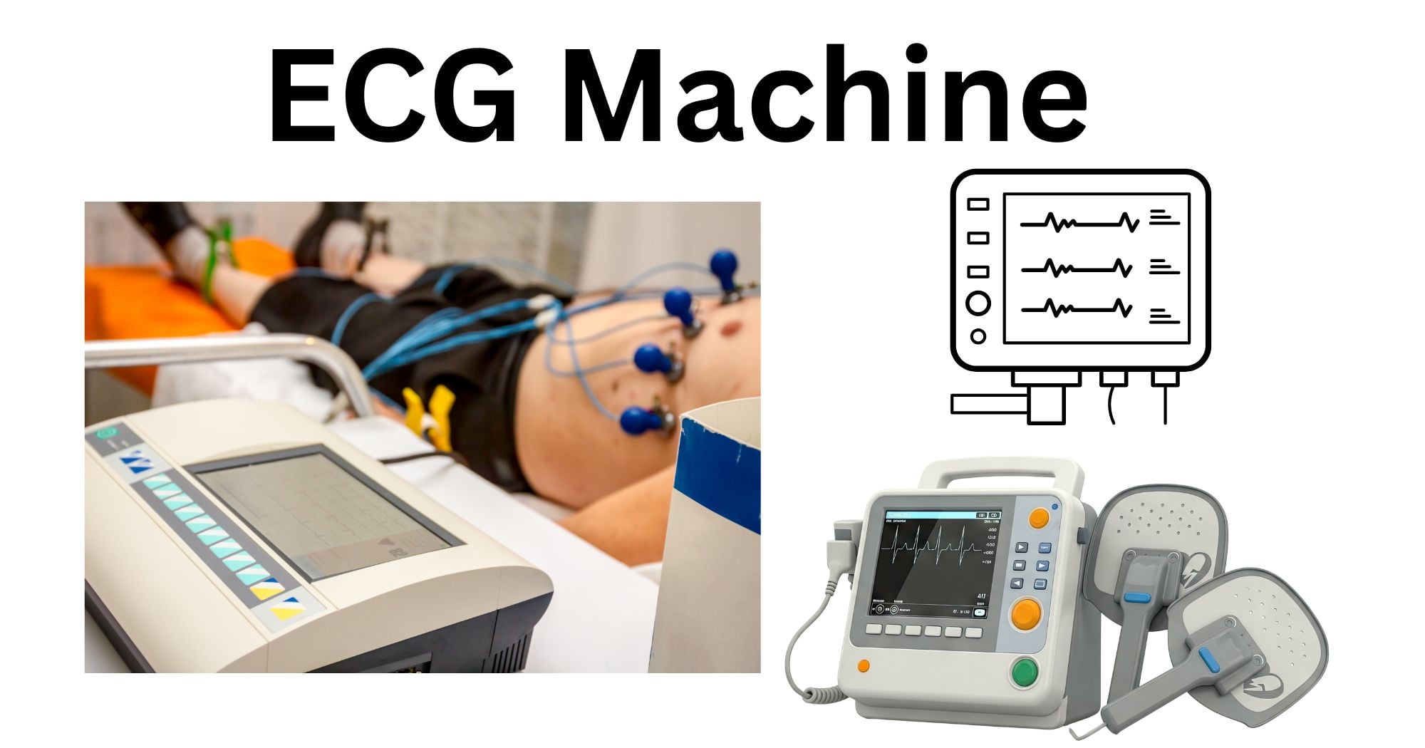
ECG machines are the most commonly used cardiac diagnostic test worldwide. They are non-invasive and painless, and provide critical information about heart rate, rhythm, conduction pathways, chamber size, and myocardial integrity.
Principle and Working of an ECG Machine
The core principle relies on the heart’s electrical conduction system. The ECG captures voltage differences on the body surface resulting from the heart’s depolarization and repolarization cycles. The pacemaker of the heart, the sinoatrial (SA) node, generates spontaneous depolarization. This propagates through the atrium, atrioventricular (AV) node, bundle of His, and Purkinje fibers to coordinate ventricular contraction. These ionic currents create measurable potential differences on the skin. The electrodes detect these potentials. The machine amplifies the weak signals, filters the noise, and records them. The result is converted from analog to digital in modern devices, which enables digital processing, storage, and automated interpretation. The standard paper speed is 25 mm/s, with 1 mV equaling 10 mm vertical deflection. When depolarization moves towards a positive electrode, it produces an upward deflection, and when it moves away, it produces a downward deflection.
Parts and Components of an ECG Machine
A typical ECG machine is composed of the following components:
- Electrodes: The electrodes are conductive pads (silver/ silver chloride) that interface with the skin.
- Lead wires and cables: These connect the electrodes to the main unit. They are often color-coded and labeled for correct placement.
- Amplifier and filter system: It multiplies, boosts, and cleans signals.
- Analog to Digital converter (ADC): This converts the analog signals.
- Display and recorder: There is a high-resolution LCD screen for real-time viewing and a thermal printer or digital storage.
- Microprocessor: It handles analysis and storage.
- Power supply: Power is provided by a battery or AC, with isolation circuits to protect the patient from electrical shock.
- User interface: Keyboard for patient data entry and settings.
Types of ECG Machines (Resting, Holter, Stress Test, and Event Monitors)
ECG machines can be classified into several categories based on their intended use and duration of recording.
Resting ECG: A resting ECG is a standard 12-lead ECG machine that captures the heart’s electrical activity over a brief period of 10-15 seconds while the patient lies supine. It is a standard instrument commonly found in hospitals and clinics, and it remains the gold standard for evaluating cardiac rhythm, ischemia, infarction, and conduction abnormalities.
Holter Monitor: This is a portable device that continuously records ECG signals over 24 – 48 hours using 3-12 leads. It captures intermittent events during daily activities. It is valuable for detecting transient arrhythmias and silent ischemia that might be missed by a resting ECG.
Stress Test ECG (Exercise ECG): It records the cardiac activity of the patient while they walk on a treadmill or bicycle to detect ischemia under stress. It is often combined with imaging.
Event Monitors: These are patient-activated or auto-triggered devices used over weeks or months. They capture ECG segments when the patient experiences symptoms or when preset arrhythmia criteria are met.
ECG Leads and Electrode Placement
A standard 12- lead ECG uses 10 electrodes to derive 12 different electrical views of the heart: 3 bipolar limb leads (I, II, III), 3 augmented unipolar limb leads (aVR, aVL, aVF), and 6 precordial leads (V1 – V6). The three limb leads, first described by Einthoven, form Einthoven’s triangle and measure potential differences between the limbs: lead I between the right and left arms, lead II between the right arm and left leg, and lead III between the left arm and left leg. The three augmented limb leads, aVR, aVL, and aVF, are derived by machine electronics rather than measured directly.
The six precordial leads, V1 to V6, are placed across the chest wall in specific intercostal spaces:
V1 in the fourth intercostal space to the right of the sternum
V2 in the fourth intercostal space to the left of the sternum
V3 midway between V2 and V4
V4 in the fifth intercostal space at the midclavicular line
V5 is at the same horizontal level as V4 at the anterior axillary line
V6 is at the same level as the midaxillary line.
Correct electrode placement is critical because misplacement distorts waveforms and diagnosis.
How to Perform an ECG Test Using an ECG Machine
Performing a standard 12- lead ECG follows a systematic protocol:
- Explain the procedure to the patient and obtain consent.
- Position the patient in the supine position with the chest exposed and instruct the patient to remain still and breathe normally.
- Prepare skin by cleaning with alcohol and shaving if hairy.
- Apply electrodes firmly and in the correct position.
- Enter patient demographic data.
- Ensure good signal quality.
- Record a 10- second rhythm strip plus a 12-lead.
- Remove electrodes carefully and document findings.
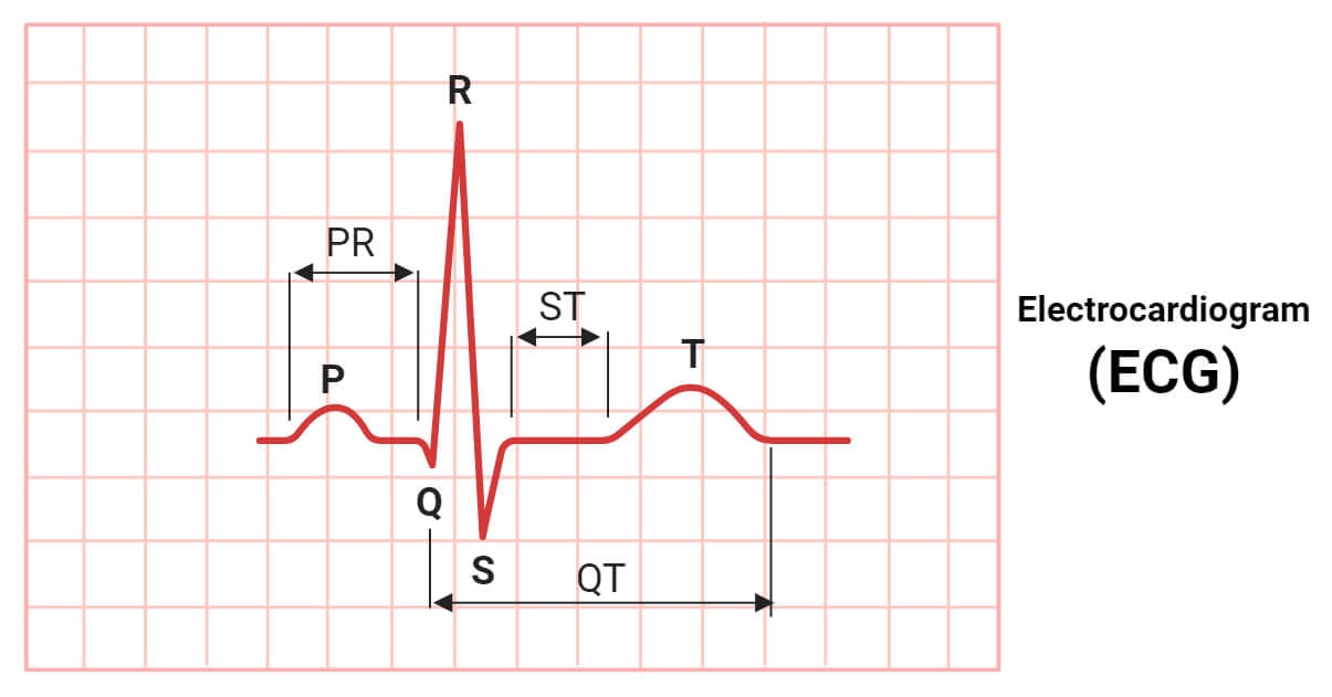
Interpretation of ECG Readings (Waves, Intervals, and Heart Rhythm Basics)
ECG interpretation is done by systematic analysis of the waveforms, intervals, and rhythm. The key components are:
- P wave: Atrial depolarization (normal < 120 ms)
- QRS complex: Ventricular repolarization (normal < 120 ms).
- T wave: Ventricular repolarization.
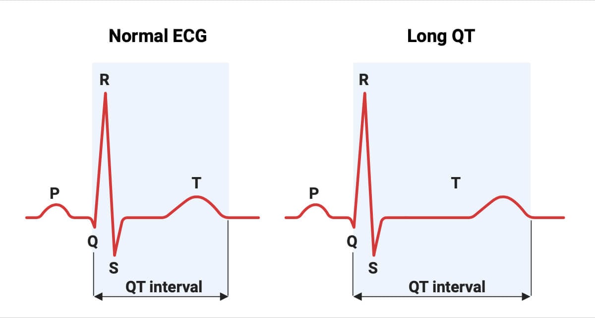
- Intervals: The PR interval is measured from the beginning of the P wave to the beginning of the QRS complex. It reflects the time required for the impulse to travel from the SA node through the AV node to the ventricles. Ranges from 0.12 to 0.20 seconds. The QT interval is measured from the QRS onset to the T wave end. It varies with heart rate. Rate- corrected QTc < 440-460 ms.
- Sections: ST (ischemia evaluation).
- Normal sinus rhythm: Regular, rate: 60 – 100 bpm, upright P in II, QRS follows each P.
- Abnormalities include ST elevation, prolonged QT, or fibrillation waves.
Applications of ECG Machines
ECG machines have a broad range of clinical applications, such as:
- Diagnosis: They are critical for diagnosing cardiac arrhythmias, including atrial fibrillation, supraventricular tachycardias, ventricular tachycardia, and bradyarrhythmias. They also detect electrolyte disturbances such as hyperkalemia and hypokalemia.
- Perioperative monitoring and preoperative risk assessment
- Evaluate the functionality of implanted cardiac devices
- Sports screening
- Research uses include drug trials and population studies
- Wearables expand screening for silent atrial fibrillation in stroke prevention
Advantages of ECG Machines
- Non-invasive, quick, inexpensive, and widely available.
- No radiation exposure, so safe for patients of all ages and pregnant women.
- Rapid (completed under 10 minutes)
- High diagnostic yield for conditions involving dysfunction of the heart.
- Portable options enable remote monitoring.
Limitations of ECG Machines
- Technical factors such as improper electrode placement, patient movement, and electrical interference may produce artifacts mimicking the pathology.
- Limited sensitivity for some conditions.
- Snapshot results, which may miss intermittent issues.
- Cannot directly assess mechanical function (complemented with echo).
Maintenance, Calibration, and Safety Considerations for ECG Machines
Regular maintenance and calibration are essential for ensuring ECG machines are accurate and safe for patients. The IEC 60601-2-25 mandates that the internal noise level must not exceed 30 microvolts peak-to-peak within the diagnostic bandwidth.
Calibration verification involves applying a known 1 mV test signal and confirming a 10 mm deflection on the recording. The input impedance must be maintained at over 2.5 megohms, and the frequency must be verified within the 0.05 to 150 Hz diagnostic range.
Safety includes electrical isolation, grounding, and defibrillator-proof design. Cables and lead wires must be inspected routinely for fraying or exposed conductors. Electrodes must be stored in sealed packages to prevent gel desiccation, and thermal printers require periodic cleaning to maintain tracing quality.
Regulatory Standards and Guidelines for ECG Machines
ECG machines are classified as Class II medical devices by the FDA and are subject to rigorous performance and safety standards.
The International Electrotechnical Commission has three particular standards: IEC 60601-2-25 (for diagnostic electrocardiographs), IEC 60601-2-27 (for ECG monitoring equipment), and IEC 60601-2-47 (for ambulatory ECG systems). These are harmonized with the Association for the Advancement of Medical Instrumentation (AAMI) standards like EC11, EC13, and EC38. The AHA/ ACC/ HRS provides clinical guidelines on recording and interpretation.
Conclusion
The ECG machine is a critical tool in cardiovascular medicine that has evolved from Einthoven’s string galvanometer to the modern digital system of today. It is a non-invasive, non-toxic, and safe method for translating subtle electrical signals of the beating heart into waves. So, a proper understanding of the technology, its handling, calibration, and maintenance is necessary for clinicians and technicians who use it. ECGs continue to bridge clinical needs with precision diagnostics and will continue to do so in the future.
References
- Sattar, Y., & Chhabra, L. (2026). Electrocardiogram. In StatPearls. StatPearls Publishing. http://www.ncbi.nlm.nih.gov/books/NBK549803/
- AlGhatrif, M., & Lindsay, J. (2012). A brief review: History to understand fundamentals of electrocardiography. Journal of Community Hospital Internal Medicine Perspectives, 2(1), 10.3402/jchimp. v2i1.14383. https://doi.org/10.3402/jchimp.v2i1.14383
- Kligfield, P., Gettes, L. S., Bailey, J. J., Childers, R., Deal, B. J., Hancock, E. W., van Herpen, G., Kors, J. A., Macfarlane, P., Mirvis, D. M., Pahlm, O., Rautaharju, P., & Wagner, G. S. (2007). Recommendations for the Standardization and Interpretation of the Electrocardiogram. Circulation, 115(10), 1306–1324. https://doi.org/10.1161/CIRCULATIONAHA.106.180200
- Petty, B. G. (2020). Components of the Electrocardiogram: The Normal Tracing. In B. G. Petty (Ed.), Basic Electrocardiography (pp. 1–17). Springer International Publishing. https://doi.org/10.1007/978-3-030-32886-3_1
- Friedman, P. A. (2024). The Electrocardiogram at 100 Years: History and Future. Circulation, 149(6), 411–413. https://doi.org/10.1161/CIRCULATIONAHA.123.065489
- Salam, A. M. (2019). The Invention of Electrocardiography Machine. Heart Views : The Official Journal of the Gulf Heart Association, 20(4), 181–183. https://doi.org/10.4103/HEARTVIEWS.HEARTVIEWS_102_19
- The ECG leads: Electrodes, limb leads, chest (precordial) leads and the 12-Lead ECG. (n.d.). The Cardiovascular. Retrieved from https://ecgwaves.com/topic/ekg-ecg-leads-electrodes-systems-limb-chest-precordial/
- Cadogan, M. (2021, May 20). ECG Lead positioning. Life in the Fast Lane • LITFL. https://litfl.com/ecg-lead-positioning/
- Electrocardiogram. (2025, September 15). https://www.hopkinsmedicine.org/health/treatment-tests-and-therapies/electrocardiogram
- Crawford, M. H., Bernstein, S. J., Deedwania, P. C., DiMarco, J. P., Ferrick, K. J., Garson, A., Green, L. A., Greene, H. L., Silka, M. J., Stone, P. H., Tracy, C. M., Gibbons, R. J., Alpert, J. S., Eagle, K. A., Gardner, T. J., Gregoratos, G., Russell, R. O., Ryan, T. H., & Smith, S. C. (1999). ACC/AHA Guidelines for Ambulatory Electrocardiography. A report of the American College of Cardiology/American Heart Association Task Force on Practice Guidelines (Committee to Revise the Guidelines for Ambulatory Electrocardiography). Developed in collaboration with the North American Society for Pacing and Electrophysiology. Journal of the American College of Cardiology, 34(3), 912–948. https://doi.org/10.1016/s0735-1097(99)00354-x
