
The skin, the human body’s largest organ, serves as a physical and immunological barrier to the external environment. It is home to a diverse and complex variety of innate and adaptive immune functions. Despite the skin’s potent immune system, it still encourages colonization by microorganisms. The skin’s large surface area is colonized by diverse microbial communities, including bacteria, viruses, fungi, and Demodex mites.
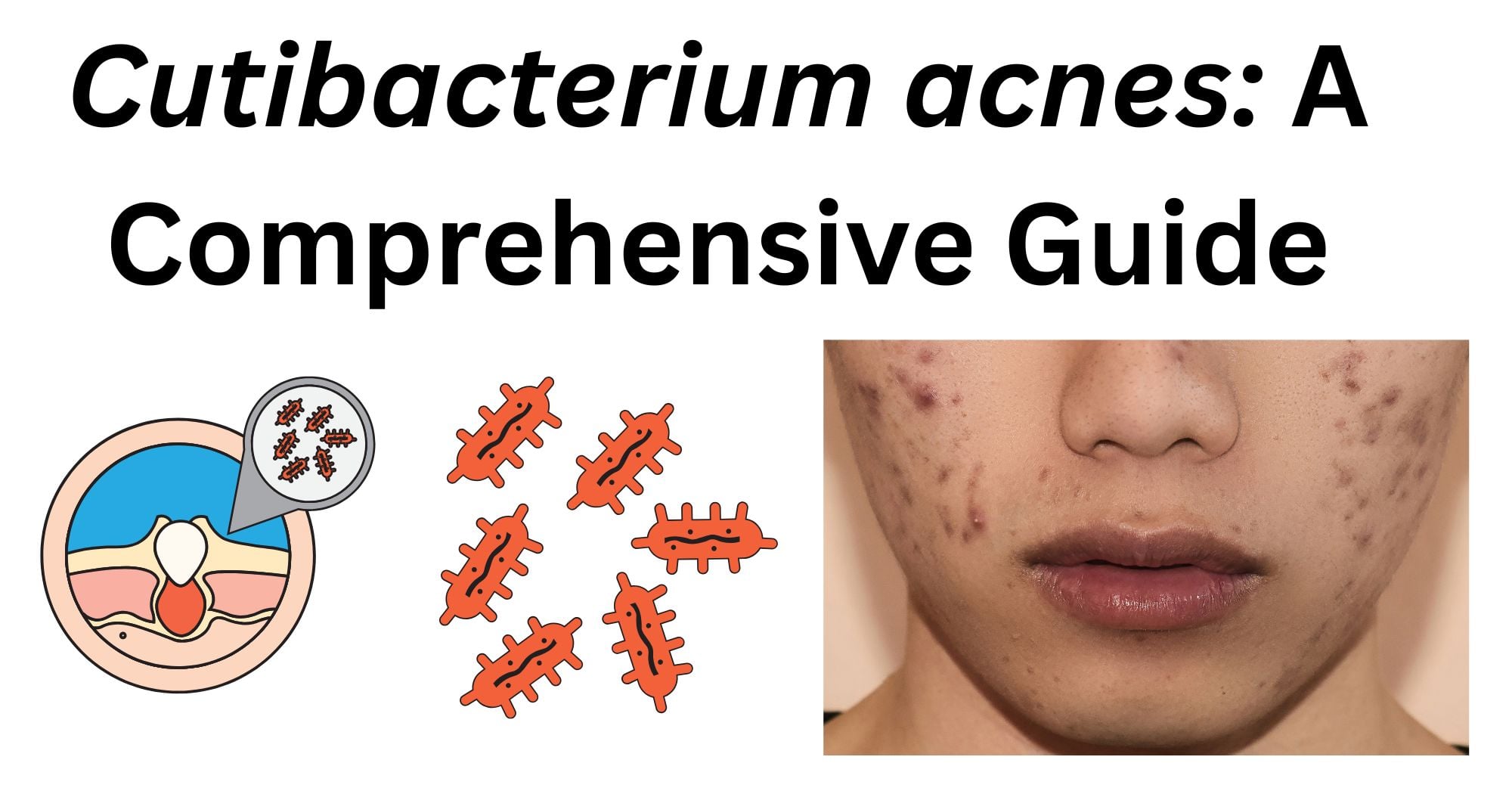
These microorganisms and their genetic material together make up the skin microbiome. One microorganism in the skin microbiome is Cutibacterium acnes, previously known as Propionibacterium acnes. It is mainly found on the skin and in the gastrointestinal tract. Acne vulgaris is one of the most common skin diseases. Microbial involvement is considered one of the main mechanisms for the development of acne, although the etiology and pathogenesis of acne are still unclear.
However, it has been shown that rather than C. acnes species as a whole, it is the loss of microbial diversity in the skin and the imbalance among C. acnes phylotypes that bring about acne.
Classification of Cutibacterium acnes
Kingdom: Bacteria
Phylum: Actinomycetota
Class: Actinomycetia
Order: Propionibacteriales
Family: Propionibacteriaceae
Genus: Cutibacterium
Species: Cutibacterium acnes
Following its isolation, Cutibacterium acnes was first included in the genus Bacillus as Bacillus acnes, and then in the genus Corynebacterium as Corynebacterium acnes. Later, it was grouped into the genus due to its ability to produce propionic acid during its anaerobic catabolism. Subsequently, it was renamed to Cutibacterium. Genus Cutibacterium belongs to a branch of Actinobacteria and can be split into two groups: “classic or dairy” species and other “skin” species mostly found on the surface of human skin.
Morphology of Cutibacterium acnes
Cutibacterium acnes is a commensal lipophilic gram-positive bacterium. It is described as diphtheroid or coryneform because it is a rod-shaped and slightly curved bacterium with a width of 0.4 to 0.7µm. Anaerobic bacteria are characterized by their inability to grow on solid media in the presence of atmospheric oxygen.
The organism is, however, considered an aerotolerant anaerobe because it possesses enzymatic systems able to detoxify oxygen, allowing it to be sustained on the surface of the skin.
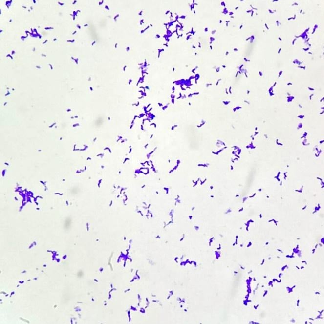
Under Gram staining, Cutibacterium acnes is observed as gram-positive, violet purple-coloured cells due to its thick peptidoglycan layer in the cell wall. These bacteria occur in singles, in pairs, or in short chains and sometimes may form irregular clusters in samples from sebaceous-rich tissues. C. acnes lack flagella and are hence non-motile. This lack of flagella is a distinguishing characteristic of the microorganism that separates it from other gram-positive rods with motility-associated structures.
While many strains are capable of producing extracellular polymeric substances that support biofilm formation, C. acnes does not form spores.
Microscopy of Cutibacterium acnes
When observed under a light microscope, the organism is seen as a small gram-positive bacillus that has retained the crystal violet and appears purple-coloured due to the thick layer of peptidoglycan layer in its cell wall.
On microscopy of clinical smears, C. acnes is often seen as short rods or diphtheroid-like cells. These cells have either an irregular form or a pleomorphic form depending on the growth environment and specimen type. Cutibacterium acnes is best visualized after anaerobic incubation. When the organism is incubated in anaerobic conditions, they retain the typical rod morphology. Depending on the incubation period and amount of nutrients available for the growing bacteria, the cells may show slight pleomorphism.
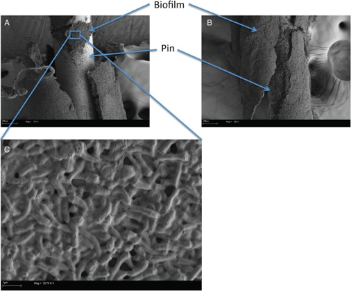
Microscopy may reveal compact bacterial masses embedded in the biofilm-like structures during acne vulgaris or implant-associated infections, which are pathological conditions.
Cultural and Growth Characteristics of Cutibacterium acnes
Cutibacterium acnes, formerly known as Propionibacterium species, is a commensal Gram-positive skin bacterium that can cause implant-associated infections. Acne vulgaris (commonly called acne) is one of the most common skin diseases, with a prevalence of up to 85% of teenagers and 11% of adults. Although the etiology and pathogenesis of acne are still unclear, microbial involvement is considered one of the main mechanisms contributing to the development of acne.
Cutibacterium acnes are slow-growing anaerobic bacteria that are also aerotolerant. They exhibit distinct cultural characteristics under laboratory conditions. Although C. acnes can tolerate a limited amount of oxygen due to its metabolic adaptations, its optimal growth occurs under an anaerobic or microaerophilic environment.
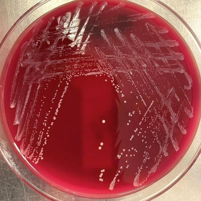
Since they are slow growers, their colonies become visible only after around 5-10 days, and this prolonged incubation requirement is one of their key characteristics. In some of the clinical contexts, this bacterium can take as long as 14-21 days for optimal isolation. This often causes the C. acnes to remain undiagnosed due to insufficient culture duration.
https://www.pathologyoutlines.com/topic/microcutibacteriumacne.html
Unlike other gram-positive bacteria, C. acnes has a unique cell wall and envelope that contain phosphatidylinositol, triglycerides, and many other common lipids. The cell wall of C. acnes consists of peptidoglycan (PNG), but of a type different from other gram-positive bacteria, in that the peptide chain contains the L-alanine, D-alanine, and D-diaminopimelic acid.
Small, round, smooth, and opaque colonies that are white to grayish in colour are produced by C. acnes on solid media like Blood Agar when cultured in anaerobic conditions. These organisms are seen to be best growing in enriched media and under anaerobic conditions, and form visible colonies after an extended incubation period. C. acnes is also observed to be able to grow in low-nutrient, low-oxygen environments persistently. They are also capable of forming biofilms, which directly enhances their growth behaviour by allowing these organisms to survive in the protected microenvironments and resist both antimicrobial agents and host immune responses.
Epidemiology of Cutibacterium acnes
Acne vulgaris (acne) is a common dermatological condition worldwide, affecting approximately 79% to 95% of people, and is a chronic inflammatory disease caused by the proliferation of Cutibacterium acnes due to hyperkeratotic abnormalities and increased sebum secretion. This disease may begin during adolescence and young adulthood. It affects both sexes, although females tend to develop acne more frequently.
Cutibacterium acnes is a bacterium that thrives in the normal human skin microbiota, which helps to maintain and support the natural microbial balance of the skin. However, under certain conditions, it can also substantially change its local environment and cause diseases.
The presence of C. acnes on human skin is nearly universal. The bacteria colonize the skin shortly after birth, but stable colonization develops during the 1-3 years preceding puberty. The bacterial count then rises dramatically from fewer than 10 cm2 to approximately 106 per cm2 on the face and upper thorax. C. acnes are seen to colonize lipid-rich, high-sebum areas, including the nose, shoulders, back, and upper chest area.
C. acnes are comprised of several distinct phylotypes and have been classified into types I, IA, IB, IC, and type III. IA1 is the most common phylotype isolated from acne patients, which accounts for about 71.4% of the strains, followed by IA2 and type III. Phylotype IA1 is also the most prevalent in both healthy skin and in implant-associated infections globally. However, the bacterium itself is present in virtually all human populations worldwide.
Pathogenesis of Cutibacterium acnes
The skin commensal Propionibacterium acnes, recently renamed Cutibacterium acnes along with the other major pathophysiological factors of increased seborrhea, hyperkeratinization of the pilosebaceous unit, and inflammation, has long been implicated in the pathogenesis of acne. Recent advances have contributed to our understanding of the role of P. acnes in acne. They are ubiquitous organisms present in skin flora and are generally found in sebaceous glands. They are notably slow-growing, facultatively anaerobic, and non-spore-forming.
Techniques such as prolonged agar cultures for 14 days, as well as tissue sonication, have increased the detection of these bacteria. More recent work recognizes that C. acnes is a common pathogen and that prolonged culture duration is required, with some investigators advocating culture incubation lengths as long as 21 days in order to increase the yield of positive cultures.
However, extended culture duration may delay a diagnosis and treatment of infection, as well as lead to culture contamination and false-positive growth, thus creating a diagnostic challenge.
The pathogenesis of Cutibacterium acnes is multifactorial, involving biofilm formation, host immune activation, and phylotype-specific virulence. Cutibacterium is a near-ubiquitous commensal but transitions to an opportunistic pathogen under specific conditions, particularly when in the presence of implanted medical devices or even within the lipid-rich follicular environment.
The central mechanism is biofilm formation. C. acnes produces an extracellular matrix that adheres to abiotic surfaces and protects the bacterium from antibiotics and host immune cells. The composition of the biofilm changes depending on the material, such as the infected implant material. The biofilm-mediated persistence explains the indolent, chronic nature of C. acnes implant infections that often present months to years after surgery.
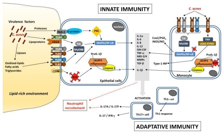
The tissue damage in acne vulgaris is driven by host inflammatory responses. The key determinant of pathogenicity in C. acnes is its phylotypic diversity. C. acnes components activate Toll-like receptors on immune cells, triggering pro-inflammatory cytokines (IL-1, IL-6, TNF-α). Different phylotypes induce distinct immune profiles: acne-associated type IA strains stimulate high levels of IFN-γ and IL-17, while healthy skin phylotypes induce the anti-inflammatory IL-10.
Virulence Factors of Cutibacterium acnes
Phylogenetic studies have shown that acquired DNA sequences and bacterial immune elements may have roles in determining the virulence properties of C. acnes strains. Biochemical, transcriptomic, and proteomic analyses demonstrated that C. acnes phylotypes exhibit differences in inflammatory potential and expression of various putative virulence factors that may explain their distinct involvement in acne disease.
These factors include neuraminidase, lipase, polyunsaturated fatty acid isomerase, and heat shock proteins. Host-interacting factors such as CAMP factors, hemolysins, and dermatan sulphate-binding adhesins have also been identified as possible pathogenic factors.

CAMP factors
The five CAMP factors encoded by the genome of all C. acnes strains are membrane pore-forming toxins that act as host tissue degradation enzymes. These secretory proteins are potentially cytotoxic for keratinocytes and macrophages, and their activation may result in skin inflammation. Recent in vitro findings indicated that CAMP1 may be involved in C. acnes virulence by interacting directly with TLR2, thus amplifying the inflammatory response. CAMP1 factor genes were found to be most strongly expressed in types IB and II, while CAMP2 factor was detected in greater amounts in IA isolates.
Porphyrins
Porphyrins are produced by C. acnes and might contribute to the perifollicular inflammatory reaction during acne development. Their ability to generate singlet oxygen from oxygen under ultraviolet exposure might enhance the production of cytotoxic substances by oxidation processes, such as squalene peroxide, a proinflammatory lipid. They can also stimulate the expression of keratinocyte-derived interleukin-8 and prostaglandin E2, which are mediators of inflammatory and immune responses.
Hyaluronate Lyase
Recently, the hyaluronate lyase has been reported with different gene alleles depending on C. acnes phylotypes. Hyaluronate lyase, along with other enzymes capable of destroying components of the dermal and epidermal cellular matrix, such as proteins, hyaluronic acid, and other glycosaminoglycans, may indeed promote the spread of inflammation during acne development.
Polyunsaturated Fatty Acid Isomerase
The PUFA isomerase from C. acnes is a yellow 424-residue monomeric protein that can catalyze the isomerization of conjugated linoleic acid (CLA). CLA and its isomers regulate several functions in humans and are present in low levels in food. Six PUFAs have been identified in C. acnes and appear to be good candidates for the development of a recombinant isomerase capable of isomerizing. This enzyme has been identified in C. acnes strains, but its role and pattern of expression in the various phylotypes remain unknown.
Other virulence factors
The acne-associated phylotype IA1 also contains a novel plasmid with a tight adhesion locus and two unique genomic islands that comprise genes supposed to enhance virulence through increased bacterial adhesion and host immune response. A correlation between the severity of acne and lipase activity has been shown with the C. acnes phylotype I that produces higher quantities of propionic acid and butyric acids than other C. acnes biotypes, and that predominates in isolates from most severe acne skins. A recent proteome analysis of human sebaceous follicle infundibula extracted from healthy and acne-affected skin revealed at least 12 putative lipases, but only two (GehA and GehB) possess a signal peptide for secretion. It is possible that other lipases produced by different C. acnes phylotypes play distinctive roles in regard to health and disease, but this hypothesis needs further investigation.
Clinical Manifestations of Cutibacterium acnes
General symptoms such as fever are relatively rare. These symptoms tend to develop gradually, contributing to the insidious nature of C. acnes infections. C. acnes is known to cause surgical infections, prosthetic joint infections, and infections of implanted devices, along with the most common manifestation, acne vulgaris.
Acne Vulgaris
It is the most common clinical manifestation of C. acnes for which one of the main causes is the overactivity of all glands, which encourage bacteria in the skin to multiply. This bacterium uses the oil gland secretions, sebum, as a source of energy. C. acnes multiply in skin with an oily build-up, leading to the inflamed spots characteristic of acne.
Other skin-related diseases associated with C. acnes
C. acnes is suspected to be involved in other skin diseases. The disease progressive macular hypopigmentation (PMH) is characterized by non-scaly hypopigmented skin areas that are visible in the sebaceous areas; often, the lower back skin is affected.
C. acnes might also play a role in acne fulminans, a rare, severe form of inflammatory acne associated with painful ulceration and, in some cases, with systemic signs.
Non-skin diseases associated with C. acnes
C. acnes and implant-associated infections
More studies report the detection of C. acnes in implant-associated infections. It seems now that C. acnes is more often found in IAI-associated specimens than two decades ago. The reason for this increase is most likely related to the changed procedures and diagnostic tools. One important change is the introduction of sonication of removed periprosthetic tissue specimens or removed medical devices before microbial cultivation. This technique is nowadays more often used in everyday clinical microbiology practice in many hospitals and has been shown to increase the bacterial recovery from specimens.
Prosthetic joint infections
C. acnes is associated with around 10% of all prosthetic joint infections (PJIs), being more frequently isolated in late-chronic infections. The shoulder is the most frequent site of isolation, probably due to the greater rate of colonization in the axillary region than in the hip or knee. C. acnes is also believed to be one of the main opportunistic pathogens involved in latent postoperative infections in spinal instrumentation surgeries.
Cardiac device-related infections
C. acnes has also been described as a causative agent of cardiovascular device-related infections, involving prosthetic heart valves, permanent pacemakers, prosthetic valve rings, or implantable cardioverter-defibrillators.
Neurosurgical shunt infections
C. acnes is considered an emerging opportunistic pathogen in neurosurgery procedures. It may be responsible for ~15% of infections associated with shunt tubular devices, that drains the cerebrospinal fluid (CSF) from cerebral ventricles to other body sites, usually the peritoneum. Clinical symptoms in shunt infections are non-specific; the absence of fever is common.
Laboratory Diagnosis of Cutibacterium acnes
Specimen
periprosthetic tissue, synovial fluid, CSF, explanted prosthetic devices: specimens are to be transported promptly to the laboratory in sterile and anaerobic containers at room temperature
Gram staining can provide an initial clue to the presence of C. acnes. The organism appears to be Gram-positive, pleomorphic, non-spore-forming, and arranged singly or in small clusters.
Culture
Being the slow-growing and fastidious anaerobe that it is, C. acnes requires extended incubation under strict anaerobic conditions for its detection. The generation time of this microorganism is longer than that of the majority of other pathogens, which is observed to be around 5.1 hours, making prolonged culture an essential part of the identification. The incubation time should be a minimum of 6 days and held up to 14 days to optimize recovery.
Thioglycolate broth has been observed to successfully support the growth of C. acnes, including in cases of infectious keratitis wherein cultures become positive only after about 7 days of incubation.
Matrix Assisted Laser Desorption/Ionization Time-of-Flight Mass Spectrometry (MALDI-TOF MS)
There has been a great improvement in the identification of C. acnes from positive cultures upon the emergence and use of MALDI-TOF MS. It provides a fast, reliable species-level identification by analyzing bacterial protein profiles. However, confirmation by molecular methods may be required in certain cases, as the MALDI-TOF MS may misidentify some closely related Cutibacterium species like C. namnetense and C. modestum.
Molecular Methods
A powerful technique for the detection of C. acnes in clinical specimens is Polymerase Chain Reaction (PCR), in particular, the universal 16S rRNA gene PCR. This method is especially useful when cultures are negative despite strong clinical suspicion and can identify the bacterium directly from tissue or sonication fluid. Amplification of 16S rRNA sequences enables sensitive detection, even in the presence of antibiotics or when bacterial viability is compromised.
Late discoveries of positive samples after apparently aseptic implant change is an at-risk situation, usually managed by antibiotic therapy, but with late initiation and hence increased risk of failure.
Treatment of Cutibacterium acnes (Acne Vulgaris)
Topical treatments
- Benzoyl Peroxide (BP)- potent, first-line antimicrobial that kills C. acnes rapidly.
- Topical Retinoids- agents like adapalene, tretinoin, and tazarotene are essential for normalizing skin cell shedding and have anti-inflammatory properties.
- Topical Antibiotics- clindamycin or erythromycin are used for inflammatory lesions, but must always be combined with benzoyl peroxide to minimize the risk of bacterial resistance.
Systemic treatments
- Oral Antibiotics- prescribed for moderate to severe inflammatory acne when rapid control is needed; first-line options include doxycycline, minocycline, and tetracycline.
- Isotretinoin- oral retinoid that is strongly recommended for severe scarring or treatment-resistant acne.
- Hormonal therapies, for selected female patients, combined oral contraceptives, or spironolactone can be effective by reducing sebum production.
Prevention of Cutibacterium acnes
Acne prevention
- Over-the-counter (OTC) Topicals- OTC products with benzoyl peroxides are first-line treatment for mild acne; BPO kills C. acnes by releasing oxygen radicals and helps clear pores without causing bacterial resistance
- Prescription Topicals- dermatologists may prescribe a topical retinoid to prevent pores from clogging in case of more persistent cases
Surgical and hospital prevention
- Preventive Decolonization- 5% BPO cream can be applied for 5 days before surgery, and is the preferred decolonization strategy
- Surgical Site Prep- combining 3% hydrogen peroxide for 5 minutes with ChloraPrep scrub is a cost-effective protocol to reduce bacterial loads.
- Antibiotic Prophylaxis- intravenous cefazolin is the prophylactic agent of choice during shoulder arthroplasty. Applying vancomycin powder deep to the surgical wound closure offers an additional local defense.
References
- Boisrenoult, P. (2018). Cutibacterium acnes prosthetic joint infection: Diagnosis and treatment. Orthopaedics & Traumatology: Surgery & Research, 104(1S), S19–S24. https://doi.org/10.1016/j.otsr.2017.05.030
- Dréno, B. (2016). Bacteriological resistance in acne: A call to action. European Journal of Dermatology, 26(2), 127–132. https://doi.org/10.1684/ejd.2015.2685
- Dréno, B., Pécastaings, S., Corvec, S., Veraldi, S., Khammari, A., & Roques, C. (2018). Cutibacterium acnes (Propionibacterium acnes) and acne vulgaris: a brief look at the latest updates. Journal of the European Academy of Dermatology and Venereology, 32(Suppl. 2), 5–14. https://doi.org/10.1111/jdv.15043
- Fitz-Gerald, P. T., Stamer, D. K., Wyles, C. C., & Houdek, M. T. (2019). Reduced time to positive Cutibacterium acnes culture utilizing a novel incubation technique: A retrospective cohort study. Journal of Shoulder and Elbow Arthroplasty, 3, 2471549219840823. https://doi.org/10.1177/2471549219840823
- Jeon, J. H., & Lee, J. W. (2019). Epidemiological survey of Cutibacterium acnes involved in acne in healthy individuals. Journal of the Japan Society of Acne Research, 68(2), 339–346. https://www.jstage.jst.go.jp/article/jamt/68/2/68_18-91/_article/-char/en
- Mayslich, C., Grange, P. A., & Dupin, N. (2021). Cutibacterium acnes as an opportunistic pathogen: An update of its virulence-associated factors. Microorganisms, 9(2), 303. https://doi.org/10.3390/microorganisms9020303
- Mohammed, E. R., Baqer, L. K., & Dhaher, S. A. (2025). Isolation and prevalence of Cutibacterium acnes phylotypes among acne patients. Romanian Journal of Medical Practice, 20(4), 374–380. https://doi.org/10.37897/RJMP.2025.4.6
- Niedźwiedzka, A., Mękal, M. P., Borzęcka, M., & Płatek, C. (2024). The role of the skin microbiome in acne: Challenges and future therapeutic opportunities. International Journal of Molecular Sciences, 25(21), 11422. https://doi.org/10.3390/ijms252111422
- Parente, J. N., Ormond, D. T., Cherian, N. J., & Miller, M. D. (2026). Cutibacterium acnes culture isolation following total hip and total knee arthroplasty. Antibiotics, 15(2), 165. https://doi.org/10.3390/antibiotics15020165
- Platsidaki, E., & Dessinioti, C. (2018). Recent advances in understanding Propionibacterium acnes (Cutibacterium acnes) in acne. F1000Research, 7, F1000 Faculty Rev-1953. https://doi.org/10.12688/f1000research.15659.1
- Pu, M., & Garrett, E. M. (2023, November 10). Cutibacterium acnes. PathologyOutlines.com. https://www.pathologyoutlines.com/topic/microcutibacteriumacne.html
- Sanford, J. A., & Gallo, R. L. (2013). Functions of the skin microbiota in health and disease. Seminars in Immunology, 25(5), 370–377. https://doi.org/10.1016/j.smim.2013.09.005
- Siddiqui, R., Makhlouf, Z., & Khan, N. A. (2022). The increasing importance of the gut microbiome in acne vulgaris. Folia Microbiologica, 67(6), 825–835. https://doi.org/10.1007/s12223-022-00982-5
- Tomida, S., Nguyen, L., Chiu, B. H., Liu, J., Sodergren, E., Weinstock, G. M., & Li, H. (2013). Propionibacterium acnes strain populations in the human skin microbiome associated with acne. Journal of Investigative Dermatology, 133(9), 2152–2160. https://doi.org/10.1038/jid.2013.21
- Wickham Micro. (2018, July 9). Fact sheet: Cutibacterium acnes. https://wickhammicro.co.uk/knowledge-and-education/cutibacterium-acnes
- Yoon, Y. J., & Lee, J. W. (2018). Comparative lipidomic profiling of the human commensal bacterium Propionibacterium acnes and its extracellular vesicles. RSC Advances, 8(27), 15241–15247. https://doi.org/10.1039/c7ra13769a
